


Anass Mehedra*, Bensalah Nasreddine, Amine Slaoui
Urology Department B, CHU Ibn Sina, Rabat, Morocco
*Corresponding Author: Anass Mehedra, Urology Department B, CHU Ibn Sina, Rabat, Morocco.
Abstract
Calyceal diverticula are rare urological anomalies, often asymptomatic and incidentally discovered. They may predispose patients to infection, nephrolithiasis, or pain, but severe complications such as septic shock are exceedingly rare.
We present the case of a 57-year-old male with a history of hypertension, admitted in septic shock of unknown origin. Initial imaging suggested a large, possibly infected renal cyst. Despite empirical antibiotic therapy, clinical and biological response was limited. Abdominal CT confirmed a 17 cm lesion in the left kidney. Percutaneous drainage under ultrasound guidance revealed purulent material, confirming the diagnosis of an infected calyceal diverticulum.
Introduction
Calyceal diverticula are rare urological anomalies, often asymptomatic and incidentally discovered. They may predispose patients to infection, nephrolithiasis, or pain, but severe complications such as septic shock are exceedingly rare.
Case Presentation And Importance
We present the case of a 57-year-old male with a history of hypertension, admitted in septic shock of unknown origin. Initial imaging suggested a large, possibly infected renal cyst. Despite empirical antibiotic therapy, clinical and biological response was limited. Abdominal CT confirmed a 17 cm lesion in the left kidney. Percutaneous drainage under ultrasound guidance revealed purulent material, confirming the diagnosis of an infected calyceal diverticulum.
Discussion
Calyceal diverticula are congenital anomalies that may remain asymptomatic or present with recurrent urinary tract infections, hematuria, or lumbar pain. Their diagnosis often relies on imaging, particularly CT urography, which can demonstrate communication with the collecting system.
Conclusion
Infected calyceal diverticula are rare but should be included in the differential diagnosis of renal cystic lesions.
Keywords: Calyceal diverticula, Renal cyst, infection, antibiotic therapy.
Introduction
Calyceal diverticulum is a rare clinical entity, more commonly described in the pediatric population, with a prevalence estimated between 0.21% and 0.6% [1]. It represents a congenital outpouching of a renal calyx, lined by urothelium and connected to the collecting system via a narrow infundibulum. This communication allows urine to enter the diverticulum, creating a stagnant environment predisposing to infection, stone formation, and pain. However, most cases remain asymptomatic and are discovered incidentally during imaging.
Case Report
We report the case of a 57-year-old male with a medical history of hypertension on medication, who presented with a 5-month history of lumbar pain and was admitted to the emergency department in septic shock of unknown origin.
After initial hemodynamic stabilization, monitoring, and biological sampling (blood and urine cultures), empirical antibiotic therapy with ceftriaxone and amikacin was initiated. Bedside ultrasound revealed a 17 cm left renal cystic lesion with anechoic content, suggestive of an infected renal cyst.
Despite 48 hours of antibiotic treatment, there was no clinical or biological improvement. A contrast-enhanced abdominal and pelvic CT scan (excretory phase) confirmed the findings without providing further diagnostic clarity (Figures 1 and 2).
Given the poor clinical response, antibiotic therapy was escalated to imipenem. This led to modest improvement, allowing weaning off vasopressors and regaining of consciousness. However, septic parameters remained elevated.
After seven days, a urological consultation was requested. Physical examination revealed tenderness in the left lumbar region and persistent fever. A percutaneous ultrasound-guided drainage was performed, yielding purulent material. Bacteriological culture identified Escherichia coli, confirming the diagnosis of an infected calyceal diverticulum (Figures 3 and 4).

Figure 1: Coronal section from an abdominal CT scan in the arterial phase showing a 17 cm renal cyst with contrast-enhancing wall and hypodense content.
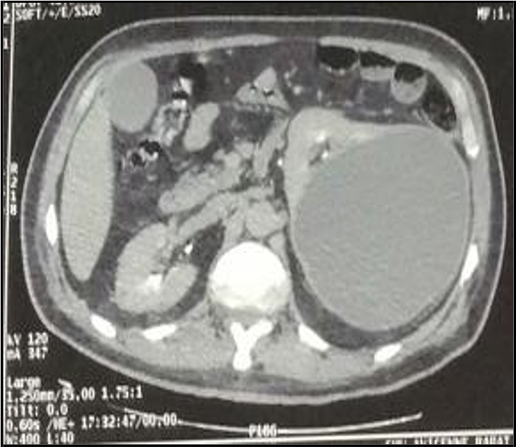
Figure 2: Coronal section from a delayed-phase abdominal CT scan showing a 17 cm renal cyst with contrast-enhancing wall and hypodense content. No communication was observed between the cyst and the pelvicalyceal system.

Figure 3: Ultrasound performed by the on-call urologist prior to nephrostomy drainage, revealing an anechoic cavity developed within the renal parenchyma.

Figure 4: Drainage fluid composed of approximately 600 cc of purulent material.
Discussion
Calyceal diverticula are congenital anomalies that may remain asymptomatic or present with recurrent urinary tract infections, hematuria, or lumbar pain. Their diagnosis often relies on imaging, particularly CT urography, which can demonstrate communication with the collecting system.
The diagnosis of a calyceal diverticulum, particularly in an acute or infectious setting, presents significant challenges. Differentiating it from a simple renal cyst requires careful imaging interpretation. CT urography during the delayed excretory phase is the preferred method, as it may reveal contrast filling of the diverticulum. Additional diagnostic clues include findings from retrograde pyelography, which is especially helpful when the infundibulum is too narrow to be visualized via CT alone. In the context of infection, wall enhancement may be seen in both cysts and diverticula, therefore limiting their specificity.
In this case, the first misidentification of the lesion as a simple infected cyst emphasizes the need for a high degree of clinical suspicion and radiologic image optimization. The patient was unable to improve on broad-spectrum antibiotics before re-evaluating the diagnosis. If there was an earlier awareness of the true diagnosis, and subsequently, the drainage procedure would have been performed sooner, the patient may not have been septic for as long. The critical takeaway is that infected calyceal diverticulum should be suspected any time an imaging cystic renal lesion is identified in a sepsis patient. Importantly, delayed diagnosis can lead to irreversible damage to the renal parenchyma. Chronic infection within a poorly draining diverticulum may result in fibrosis, abscess formation, or progressive parenchymal loss. If not adequately managed, this may be responsible for loss of renal function [2]. Early drainage, as in this case, not only reduced the risk of surgical intervention but likely preserved renal tissue viability.
The therapeutic approach to calyceal diverticulum infection is further complicated by the anatomy of the anomaly itself. The narrow infundibular neck can significantly limit the effectiveness of percutaneous drainage. Even when fluid is aspirated, residual debris or calculi may remain trapped due to impaired outflow. In such cases, a more aggressive attitude is mandatory, and definitive treatment such as percutaneous nephrolithotomy (PCNL), endoscopic fulguration, or surgical excision may be required as a second-stage procedure, once the infectious episode is adequately controlled [3].
Imaging performed after drainage, using contrast injection through the catheter or retrograde pyelography, can be useful to evaluate the patency and structure of the diverticulum.
Ultimately, this case illustrates the rare but severe consequences of infected calyceal diverticula and emphasizes the value of a multidisciplinary approach, integrating radiology, urology, and infectious disease management. Awareness and timely intervention are essential to prevent morbidity and preserve renal function.
Conclusion
Infected calyceal diverticula are rare but should be included in the differential diagnosis of renal cystic lesions associated with systemic infection. Early imaging and multidisciplinary management are crucial for a favorable outcome.
Conflicts of Interest: None
Sources of Funding: Individual
Ethical Approval:
Ethical approval was not required for this case report in accordance with the policy of CHU Ibn Sina, Rabat, Morocco. The report is based on a single patient and presents no experimental intervention. Written informed consent was obtained from the patient for publication of this case and accompanying images.
Consent
Written informed consent was obtained from the patient for publication of this case and accompanying images.
This case has been reported in line with the SCARE 2023 criteria.
References
- Sahin H, Sarioglu FC, Alaygut D, Akdogan AI, Pekcevik Y (2020) Differentiation of simple renal parenchymal cyst and calyceal diverticulum. Pediatr Int. 62(5): 615-623.
- Mehedra A, Maachi Y, Babty M, Slaoui A, Karmouni T, et al. (2025) Exploring retroperitoneal fibrosis: Insights, challenges, and treatment approaches. Urologia. 92(1): 14-20.
- Kranz J, Bartoletti R, Bruyère F, Cai T, Geerlings S, et al. (2024) European Association of Urology Guidelines on Urological Infections: Summary of the 2024 Guidelines. Eur Urol. 86(1): 27- 41.