


Vineet Banga1*, Stuti Jain2, Anju Gupta3, Sachi Gupta4
1HOD Pathology, Deptt. Of Pathology, Acharyashree Bhikshu Government Hospital, Moti Nagar, New Delhi
2Senior Resident, Deptt. Of Pathology, Acharyashree Bhikshu Government Hospital, Moti Nagar, New Delhi
3HOD Surgery, Deptt. Of Surgery, Acharyashree Bhikshu Government Hospital, Moti Nagar, New Delhi
4Junior Resident, Deptt. Of Surgery, Acharyashree Bhikshu Government Hospital, Moti Nagar, New Delhi
*Corresponding Author: Vineet Banga, HOD Pathology, Deptt. Of Pathology, Acharyashree Bhikshu Government Hospital, Moti Nagar, New Delhi, India
Abstract
Epidermal inclusion cyst (EIC) of the breast is an uncommon benign condition and is usually located in the skin layer. EIC refers to cysts that result from the proliferation and implantation of epidermal elements within a circumscribed space in the dermis. Such cysts can occur anywhere in the body, although they are more common on the face, trunk, neck, extremities, and scalp. The occurrence of EIC in the breast is rare. Lesions of such nature are frequently thought to be breast lumps, considered gynecomastia or carcinoma in male breasts, and are not included as one of the primary differential diagnoses of breast lesions. We report a case of EIC of the breast in male patients so that it should be kept as a differential diagnosis of a breast lump.
Keywords: Epidermal Inclusion Cyst, Breast, FNAC
Introduction
An epidermal cyst is a benign keratin-filled cyst of pilosebaceous origin. [1] This can occur at any site in the body, with common sites being the head and neck region, trunk, and extremities. [2] An epidermal inclusion cyst refers to a cyst that results from the proliferation and implantation of epidermal elements within a circumscribed space in the dermis. These cysts grow through the accumulation of epithelial and keratinous debris. Histopathologically, these lesions are formed by the inclusion of keratinizing squamous epithelium within the dermis resulting in a cyst filled with lamellated keratin. [3] Epidermal cyst in the breast mainly presents as a palpable lump localized primarily in the periareolar region. This study aims to do a clinical, radiological, and histopathological correlation and to evaluate the role of Fine Needle Aspiration Cytology (FNAC) in its diagnosis.
Case Report
A 52-year-old male patient presented to surgery OPD with a complaint of a lump in the Right breast for 3 years, gradually increasing in size. On examination, a 3×2 cm, non-fluctuant, firm, non-tender swelling was present beneath the skin in the periareolar area. (Figure 1) There was no pain or nipple discharge associated with the node. The patient was referred for FNAC with clinical suspicion of carcinoma breast. FNA yielded pultaceous material, and the smear shows numerous anucleate squames and nucleate benign squamous cells. There were no atypical cells. The diagnosis was given as Epidermal Inclusion Cyst. (Figure 2) Radiological correlation has been done as the breast is a rare site for Epidermal cysts. Ultrasonography revealed a well-defined heterogeneous hypoechoic lesion measuring 29×28×18 mm in the right periareolar region with posterior acoustic enhancement. (Figure 3) CT was also performed, and a well-defined subareolar cystic lesion showed homogenous density without necrosis, calcification, or abnormal vascularity. (Figure 4) Excision was performed (Figure 5,6), and was sent for histopathological examination, where the diagnosis of an epidermal cyst was confirmed. (Figure 7)
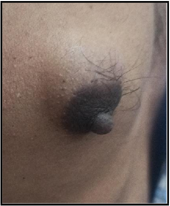
Figure 1: Lump on Right breast
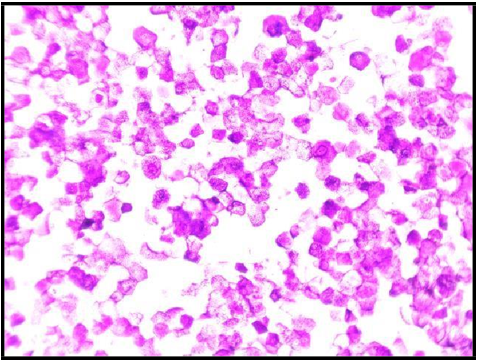
Figure 2: Giemsa stained FNAC Smears showing anucleate squames
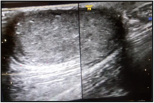
Figure 3: Ultrasound showing hypoechoic lesion in the breast
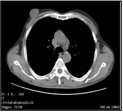
Figure 4: CT scan showing well defined lesion in the breast
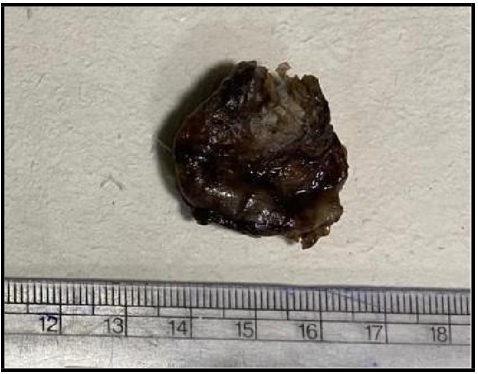
Figure 5: Gross specimen measuring 3.0 cm X 2 cm.
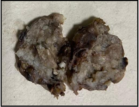
Figure 6: Cut section showing pultaceous mateial
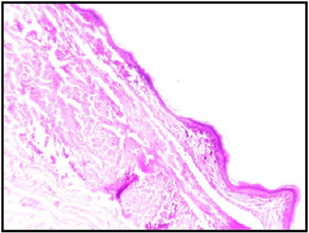
Figure 7: Microphotograph showing epithelial lining in the epidermal inclusion cyst.
(Hematoxylin & Eosin stain, X 100 Magnification)
Discussion
Gynecomastia is considered the most common breast condition affecting male patients and is a benign proliferation of ductal and stromal tissue. It is essential to consider the other differential diagnosis also. However, the epidermal cyst is a rare condition that should be viewed as a diagnosis. Epidermal cysts are primarily small, spherical, slightly compressed dome-shaped cysts with black keratin-filled punctum in the center. [4] The cyst wall is composed of stratified squamous epithelium and keratinocyte shedding from the wall resulting in the accumulation of white cheesy material with an unpleasant smell. These are usually asymptomatic and slow growing but, in a few cases, might become inflamed or secondarily infected, which may lead to pain and tenderness. [5] The possible etiology in our case can be an obstruction as there is no history of trauma or previous surgery. [6] Multiple epidermal cysts, especially in unusual locations, can be associated with a few syndromes, the most common being Gardner syndrome. Epidermal cysts in the breast can be enormous in size though it is sporadic. [3,7] These cases have higher chances of developing malignancy. [8] Singh M et al. reported six cases of EIC of the breast and concluded that it is not a rare lesion. The patients may not seek medical attention because of minor painless swelling; unless the lump increases in size or becomes painful. FNAC is confirmatory in the presence of a typical pultaceous aspirate and cytomorphological features of EIC. Thus, FNAC plays a crucial role in its diagnosis and management. Symptomatic cases should be readily excised and need histological correlation to rule out any potential complications that can arise in these cysts. [9]
Conclusion
In conclusion, the size of the breast enlargement has to be considered, and gynecomastia is the most common diagnosis, but we should consider another differential diagnosis also. EIC of the breast is rare and often mistaken as a benign or malignant condition clinically and radiologically. Therefore, FNAC plays a vital role in providing an accurate pre-operative diagnosis of EIC.
References
- Sharma S, Pujani M (2012) Epidermoid cyst of breast: A clinical and radiological dilemma resolved by FNAC. J Cytol. 29(2): 155- 156.
- Lam SY, Kasthoori JJ, Mun Ks, Rahmat K (2010) Epidermal inclusion cyst of the breast: A rare benign entity. Singapore Med J. 51(12): e191–e194.
- Lee Y, Park SG (2012) Giant sized epidermal inclusion cyst of breast initially mimicking a large fibroadenoma or phyllodes tumor. J Korean Surg soc. 83(2): 107-110.
- Fujiwara M, Nakamura Y, Ozawa T, Kitoh A, Tanaka T, et al. (2004) Multilocular giant epidermal cyst. Br J Dermatol. 151(4): 943–5.
- Swygert KE, Parrish CA, Cashman RE, Lin R, Cockerell CJ (2007) Melanoma in situ involving an epidermal inclusion (infundibular) cyst. Am J Dermatopathol. 29(6): 564–5.
- Raghupathi S, Suma S, Prakash S, Sreeramulu PN (2017) Epidermal inclusion cyst of the breast. J Surg. 5(3): 1-2.
- Meena SP, Rodha M, Badkur M, Lodha M (2020) Unusual location of subcutaneous dermoid cyst in breast: A case report. IJMSIR. 5(2): 205 – 208.
- Chiu MY, Ho ST (2007) Squamous cell carcinoma arising from an epidermal cyst. Hong Kong Med J. 13(6): 482–4.
- Singh M, Maheshwari B, Khurana N, Jain S (2012) Epidermal inclusion cyst in breast: Is it so rare?. J Cytol. 29(3): 169-172.