


Gunnar Hillerdal1, Hirsh Koyi1,2,3*
1Department of Respiratory Medicine, Gävle Hospital, Gävle, Sweden.
2Department of Oncology–Pathology, Karolinska Biomics Center, Karolinska Institutet, Stockholm, Sweden.
3Centre for Research and Development, Uppsala University/County Council of Gävleborg, Gävle, Sweden.
*Corresponding Author: Hirsh Koyi, Department of Respiratory Medicine, Gävle Hospital, Gävle, Sweden, Department of Oncology– Pathology, Karolinska Biomics Center, Karolinska Institutet, Stockholm, Sweden, Centre for Research and Development, Uppsala University/County Council of Gävleborg, Gävle, Sweden.
Introduction
Immune checkpoint inhibitors have found a place in treatment of lung cancer since some years with sometimes very good results. However, interfering with the immune system can cause a number of different side effects. Pembrolizumab is a type of targeted therapy drug called an immune checkpoint inhibitor. It is a monoclonal antibody that binds to the protein PD-1 on the surface of immune cells called T cells. It works by preventing cancer cells from suppressing the immune system [1]. We here report one unexpected reaction with this drug: an enhancement of existing tattoos.
Case Report
A 49-year-old male patient, former smoker, was referred because of shoulder pain, a left-sided pleural effusion, and enlarged mediastinal lymph nodes.
Investigated revealed a metastasizing pulmonary adenocarcinoma without any targetable mutations. Treatment was started with Carboplatin, Pemetrexed, and Pembromulizumab, and after the first four courses of chemotherapy, Pembrolizumab only, 200 mg every four weeks. After the second course of single Pembrolizumab, the patient reported that in his tattoos, some markings were shiny and standing out (Figure 1) but painful only when touched. His tumor has responded somewhat, with smaller lymph nodes and the pleural fluid disappearing. The patient was not particularly worried about the tattoo reactions but thought they were slightly improved, and we did not consider that a biopsy was warranted, nor any local treatment of them.
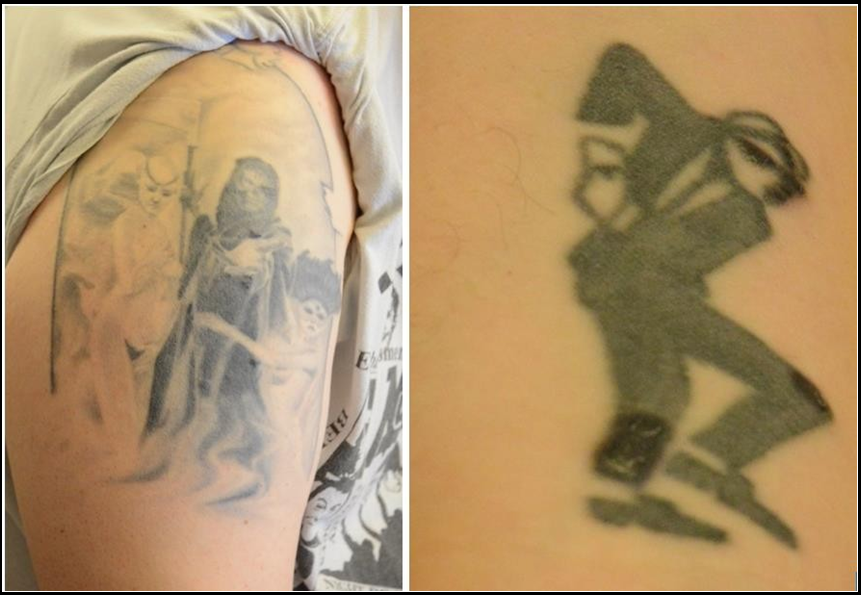
Figure 1 and 2: The darker areas are in fact standing out, making the tattoo almost three-dimensional.
Discussion
Tattoos are increasingly popular, especially among younger people [2]. Injection of tattoo pigments may alter local immune responses, creating an immunocompromised area with localized cutaneous disease [3-4].
To our knowledge, only one case of tattoo reactions in lung cancers treated with immune checkpoint inhibitors has been published [5].
Our case is very similar to this earlier publication, in which a biopsy was made and revealed a sarcoid-like reaction. Tattoo reactions in melanoma patients treated with immune therapies have been described [6]. The mechanism is unclear, but presumably, the tattoo material causes long-term, low-grade inflammation, which is enhanced by the immune system when exposed to the checkpoint inhibitor. The immune treatment might also break the immune tolerance to tattoo ink, unmasking a hypersensitivity reaction [7]. It is also well known that when a patient develops sarcoidosis, a disease that affects the immune system, former cutaneous scars can get inflamed [8].
Any biopsy of such a lesion is rarely warranted, and if done, there is a risk of deforming the tattoo picture. Treatment, if needed, is by local steroids. Several “tattoo reactions” have been described in targeted therapy for melanoma [3] but in these cases, the inflammation seems to have been more extensive.
References
- Khoja L, Butler MO, Kang SP, Ebbinghaus S, Joshua AM (2015) Pembrolizumab. J Immunother Cancer. 3: 36.
- Hogsberg T, Hutton Carlsen K, Serup J (2013) High prevalence of minor symptoms in tattoos among a young population tattooed with carbon black and organic pigments. J Eur Acad Dermat Venerol. 27(7): 846-852.
- Rucco V, Brunetti G, Puca RV, Ruocco E (2009) The immunocompromised district: A unifying concept for lymphedematous, herpes-infected and otherwise damaged sites. J Eur Acad Venerol. 23: 1364-1373.
- Ruocco V, Ruocco E, Brunetti G, Sangiuliano S, Wolf R (2011) Opportunistic localization of skin lesions on vulnerable areas. Clin Dermatol. 29(5): 483-488.
- Rousseau PM, Raimbourg J, Robert M, Dansette D, Dréno B, et al. (2019) First case of cutaneous sarcoidosis within tattoos under durvalumab. Int J Dermatol. 58(9): e168-e170.
- Kluger N (2019) Tattoo reactions associated with targeted therapies and immune checkpoint inhibitors for advanced cancers: A brief review. Dermatology. 235: 522-524.
- Rohmer E, Scrivener JN, Schissler C, Cribier B, Lenormand C (2017) Hypersensibilté retardeé aux tauotuages induites par un traitement combiné anti-BRAF.ant-Mek. Ann Demat Venerol. 144(12): S320.
- Chudomirova K, Veichikova L, Anavi B, Arnaduova M (2003) Recurrent sarcoidosis accompanying systemic sarcoidosis, J Eur Acad Dermat Venerol. 17(3): 360-1.