


Eric Curtis1, David S. Constantinescu2*, William Pavlis3, Joseph Geller2, Justin Trapana2, Brian Curtis4
1Department of Orthopaedics, University of California San Francisco Fresno, Fresno, CA, USA.
2Department of Orthopaedics, University of Miami, Miami, FL, USA.
3University of Miami Miller School of Medicine, Miami, FL, USA.
4Department of Radiology, VA National Teleradiology Program, 4150 Clement Street, San Francisco, CA, 94121, USA.
*Corresponding Author: David S. Constantinescu, Department of Orthopaedics, University of Miami, Miami, FL, USA.
Abstract
Background: The iliopsoas bursa is the largest bursa in the body, is present in the majority of people, and is located between the psoas tendon and the hip joint. Iliopsoas bursitis (IB) can be associated with hip osteoarthrosis, trauma, overuse, infection, and inflammatory arthropathies. IB may cause pain in the lower abdomen, groin, or knee, and pathologies involving the iliopsoas can cause positive psoas and obturator signs. On physical exam, IB may be confused for a hernia or an aneurysm as it may present as a mass lesion in the groin and can transmit pulsations from the nearby femoral artery. Given these nonspecific presentations, IB is an often-overlooked cause of pain.
Case Presentation: A 45-year-old, male veteran presented to the emergency room with right lower quadrant (RLQ) abdominal pain. Medical history was significant only for chronic right hip pain related to osteoarthrosis (OA). Vital signs were normal, and the exam was notable for tenderness to palpation in the right lower quadrant and inguinal region without peritoneal signs. Axial, coronal, and sagittal views on CT scan showed a lobulated fluid attenuation in the typical location of the iliopsoas bursa with an associated hip joint effusion. The diagnosis of iliopsoas bursitis explained the patient’s symptoms and the emergency room physician discharged the patient with conservative measures.
Discussion and Conclusions: Given the high morbidity and mortality associated with many causes of RLQ pain, a definitive diagnosis is aggressively pursued to ensure appropriate treatment. Musculoskeletal disorders such as iliopsoas bursitis can also be a source of RLQ pain but can be easily overlooked on CT. Medical providers interpreting imaging should be aware of the CT findings of iliopsoas bursitis as it can be a cause of RLQ pain.
Keywords: Iliopsoas Bursitis; Osteoarthritis; CT scan; Case Report
List of abbreviations: IB: iliopsoas bursitis; CT: computed tomography; RLQ: Right lower quadrant; OA: osteoarthritis.
Introduction
The iliopsoas bursa is a fluid-filled structure lined by a synovial membrane that lies between the iliopsoas muscle and the anterior aspect of the hip joint. Iliopsoas bursitis (IB) can result from several factors, such as overuse injury, septic arthritis, acute trauma, rheumatoid arthritis, and osteoarthritis [1]. The condition is characterized by distension of the bursa due to hypertrophic synovium or synovial fluid [2]. Symptoms commonly include pain in the groin that may be aggravated by flexion of the hip against resistance. The clinical presentation is similar to other disorders of the hip such as hip joint synovitis, avascular necrosis of the femoral head, and labral tears [3]. The non-specific quality of the symptoms may also lead clinicians to confuse the presentation of IB with other causes of lower abdominal or groin pain. Thus, diagnosing IB can be difficult, and imaging is commonly required to differentiate IB from other conditions, with ultrasound traditionally the most widely used method [1]. We present the case of a 45-year-old veteran with osteoarthritis of the hip presenting with RLQ pain found to have iliopsoas bursitis identified on a CT scan.
Patient history and Case presentation:
A 45-year-old, male veteran presented to the emergency room with right lower quadrant (RLQ) abdominal pain. The pain began a few days prior and was progressively intensifying with radiation from the right lower quadrant to the inguinal region. Medical history was significant only for chronic right hip pain related to osteoarthrosis (OA) from a remote injury. On presentation, he was afebrile with normal vital signs. The exam was notable for tenderness to palpation in the right lower quadrant and inguinal region. Peritoneal signs were absent and there were no palpable masses or signs of hernia. CT with the contrast of the abdomen and pelvis was performed to rule out acute appendicitis, which showed a normal appendix and findings compatible with iliopsoas bursitis.
Imaging findings:
Axial (Figure A), coronal (Figure B), and sagittal (Figure C) images from a CT of the abdomen and pelvis with intravenous contrast showed iliopsoas bursitis. In Figure A, iliopsoas bursitis, identified with an arrow, is indicated by a lobulated fluid attenuation, comparable to that found in the bladder in the image, in the typical location of the iliopsoas bursa located posterolateral to the femoral vessels (curved arrow), anterior to the hip joint, and adjacent to the iliopsoas musculotendinous junction (arrowhead). There is an associated hip joint effusion (small arrows). Subtle findings, such as these, make CT evaluation of musculoskeletal structures difficult. In Figure B, Iliopsoas bursitis (arrow) extends superiorly toward the pelvis and lateral to the femoral vessels (curved arrow). The iliopsoas musculotendinous junction (arrowhead) is slightly denser than the adjacent iliac (I) and psoas (P) muscles. In Figure C, Iliopsoas bursitis (arrow) and the adjacent psoas tendon (arrowhead) are seen coursing anterior to the hip joint.
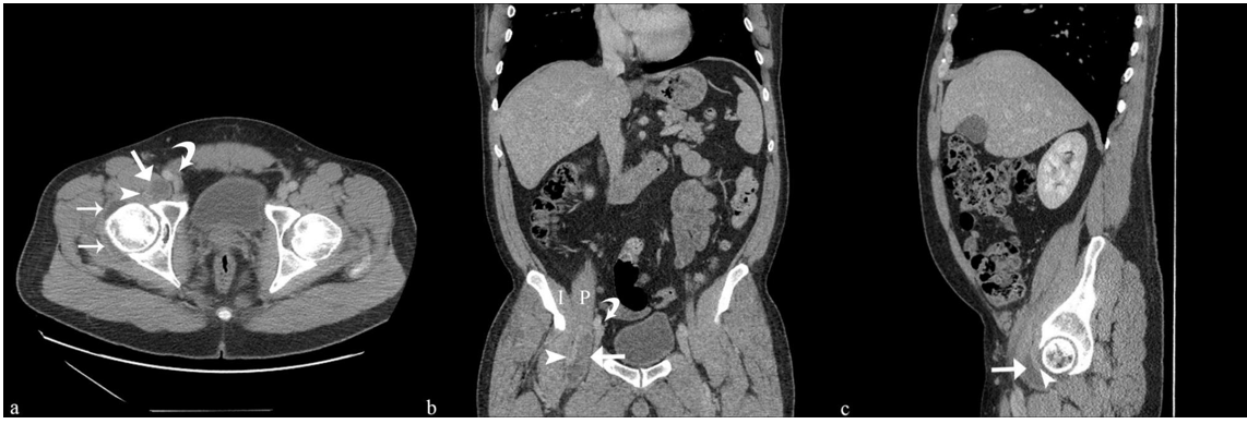
Figure A: Axial images from a CT of the abdomen and pelvis, iliopsoas bursitis, identified with an (arrow) is indicated by a lobulated fluid attenuation.
Figure B: Coronal images from a CT of the abdomen and pelvis, Iliopsoas bursitis (arrow) extends superiorly toward the pelvis and lateral to the femoral vessels (curved arrow)
Figure A: Sagittal images from a CT of the abdomen and pelvis Iliopsoas bursitis (arrow) and the adjacent psoas tendon (arrowhead) are seen coursing anterior to the hip joint.
Discussion
Acute RLQ pain is a commonly encountered complaint in the emergency setting. Given the high morbidity and mortality associated with many causes of RLQ pain, a definitive diagnosis is aggressively pursued to ensure appropriate treatment. A careful history and physical exam alone are often sufficient to identify the surgical emergencies, acute appendicitis being the most common of these. In patients with a less typical presentation, as the one presented here, adjunctive imaging is often used to help establish a diagnosis. CT is a commonly used modality in the emergency setting with high sensitivity and specificity for diagnosing acute appendicitis as well as other causes of RLQ pain including cecal diverticulitis, urolithiasis, tumors, and others. Musculoskeletal disorders can also be a source of RLQ pain but can be easily overlooked on CT. Thus, incorporating an evaluation of musculoskeletal structures into the search pattern used to interpret CT imaging of the abdomen and pelvis is necessary to not overlook pathology.
Iliopsoas disorders, such as iliopsoas bursitis, have been reported to mimic appendicitis, aneurysm, and masses [4,5]. The iliopsoas bursa is the largest bursa in the body, is present in the majority of people, and is located between the psoas tendon and the hip joint, with which it communicates in ~15 % of people [6]. Bursae are synovial lined sacs, normally containing a small amount of synovial fluid, that serves to minimize friction between adjacent musculoskeletal structures. Inflammation of bursae, or bursitis, can occur for a variety of reasons and be symptomatic. IB can be associated with hip osteoarthrosis, trauma, overuse, infection, and inflammatory arthropathies. IB may cause pain in the lower abdomen, groin, or knee (given the proximity of the bursa to the femoral nerve), and pathologies involving the iliopsoas can cause positive psoas and obturator signs. On physical exam, IB may be confused for a hernia or an aneurysm as it may present as a mass lesion in the groin and can transmit pulsations from the nearby femoral artery. Given these nonspecific presentations, IB is an often-overlooked cause of pain.
Our patient presented with acute RLQ and inguinal pain and was found on CT to have iliopsoas bursitis, most likely related to hip osteoarthrosis with a degenerative hip joint effusion. One study reported iliopsoas bursitis to affect >2 % of individuals with symptomatic Kellgren-Lawrence grade II to IV hip OA [7]. In such cases, it is hypothesized that the inflammation is due to hip joint effusion decompressing into the bursa. The iliopsoas bursa may become so distended that it extends superiorly under the inguinal ligament and displaces pelvic structures. Medical providers interpreting imaging should be aware of the CT findings of iliopsoas bursitis as it can be a cause of RLQ pain. In the case presented, the diagnosis of iliopsoas bursitis explained the patient’s symptoms and the emergency room physician discharged the patient with conservative measures.
Declarations:
Ethics Approval & Consent to Participate: The patient provided informed consent for participation in this case report.
Consent for publication: The patient provided informed consent for publication of their personal
Availability of data and materials: Data sharing is not applicable to this article as no datasets were generated or analyzed during the current study.
Competing Interests: The authors declare they have no competing interests.
Funding: Not applicable.
Authors’ contributions: EC, DC, and BC analyzed the patient data regarding patient presentation, interpretation of imaging, and outcomes. EC, DC, JG, and JT were major contributors to the development and writing of the manuscript. All authors read and approved the final manuscript.
Acknowledgments: Not applicable
References
- Johnston CA, Wiley JP, Lindsay DM, Wiseman DA (1998) Iliopsoas bursitis and tendinitis. A review. Sports Med. 25(4): 271-83.
- Corvino A, Venetucci P, Caruso M, Tarulli FR, Carpiniello M, et al. (2020) Iliopsoas bursitis: The role of diagnostic imaging in detection, differential diagnosis and treatment. Radiol Case Rep. 15(11): 2149-2152.
- Corvino A, Venetucci P, Caruso M, Tarulli FR, Carpiniello M, et al. (2020) Iliopsoas bursitis: The role of diagnostic imaging in detection, differential diagnosis and treatment. Radiol Case Rep. 15(11): 2149-2152.
- Wysoki MG, Angeid-Backman E, Izes BA (1997) Iliopsoas myositis mimicking appendicitis: MRI diagnosis. Skeletal Radiol. 26(5): 316-8.
- Peters JC, Coleman BG, Turner ML, Arger PH, Mulhern CB Jr, et al. (1980) CT evaluation of enlarged lliopsoas bursa. American Journal of Roentgenology. 135(2): 392-394.
- Van Dyke JA, Holley HC, Anderson SD (1987) Review of iliopsoas anatomy and pathology. Radiographics. 7(1): 53-84.
- Tormenta S, Sconfienza LM, Iannessi F, Bizzi E, Massafra U, et al. (2012) Prevalence study of iliopsoas bursitis in a cohort of 860 patients affected by symptomatic hip osteoarthritis. Ultrasound Med Biol. 38(8): 1352–6.