Mahmoud Omranifard M.D1, Morteza Mirzaei M.D.2, Maryam Mahabadi M.D3*, Danyal Omranifard M.D3, Majid Kalbasi Gharavi M.D2, Nadia Omranifard M.D4
1Department of Plastic and Reconstructive Surgery, Isfahan University of Medical Sciences
2Fellowship Student of Plastic and Reconstructive Surgery, Department of Plastic andReconstructive Surgery, Isfahan University of Medical Sciences
3Health and Clinical Researcher, Dr. Omranifard Clinic of Plastic and Reconstructive Surgery
4Resident student of Psychiatry, Department of Psychiatry, Shahid Beheshti University of Medical Sciences.
*Corresponding Author: Maryam Mahabadi MD, Dr. Omranifard Clinic of Plastic and Reconstructive Surgery.
Abstract
Background:
The endoscopic forehead lift is a popular surgical technique for facial rejuvenation. This study aims to evaluate the clinical and aesthetic outcomes of the three-incision endoscopic forehead lift in a cohort of patients, highlighting satisfaction levels and potential complications.
Method:
This retrospective case series included eight patients who underwent the three-incision endoscopic forehead lift between 2011 and 2022. Patient characteristics, surgical details, and aesthetic outcomes were assessed. Complications were documented, and patient satisfaction was measured using the Client Satisfaction Questionnaire (CSQ-8, v. TMS-180S).
Results:
Patients ranged in age from 35 to 65 years, with an equal distribution of males and females. Surgical scars measured between 1 and 2 cm, and the most common complication was paresthesia, affecting seven patients. Despite this, nearly all participants reported high satisfaction with their outcomes, indicating the procedure's effectiveness in enhancing facial aesthetics.
Conclusion:
The three-incision endoscopic forehead lift demonstrates significant potential for improving aesthetic outcomes with high patient satisfaction. While complications such as paresthesia may occur, the overall results support its use as an effective option for facial rejuvenation. Further research with larger sample sizes and comparative studies is recommended to strengthen these findings.
Keywords: Endoscopic forehead lift, aesthetic outcomes, clinical outcomes, three-incision method.
Introduction
Facial appearance is more than just the physical dimensions of facial features; it encompasses the emotional expressions conveyed through facial movements. These expressions serve as a primary mode of communication for conveying emotions, fostering empathy, and deeper connections among individuals. Additionally, facial expressions play a significant role in signaling characteristics like age, with lines and wrinkles reflecting a lifetime of experiences. Essentially, the face serves as a universal language, transcending cultural barriers to facilitate emotional understanding and connection, from infancy to old age, shaping how we relate to one another and perceive the world around us [1,2].
As individuals age, the wrinkles on the forehead and glabella tend to become more pronounced and static, contributing to a fixed facial appearance. This phenomenon is commonly associated with advancing age and may elicit negative perceptions regarding youthfulness or vitality [3]. Over time, a multitude of factors including genetics, skin elasticity, muscle activity, facial structure, as well as intrinsic and extrinsic influences, contribute to the formation and persistence of these lines [1, 3]. The lines that develop in the glabella region can eventually evolve into permanent wavy furrows on the forehead, commonly referred to as glabellar lines. These lines tend to worsen progressively over time, reflecting the cumulative effects of aging and environmental factors on facial appearance [5,6]. Currently, among non-surgical interventions, hyaluronic acid filler and botulinum toxin are the main materials used for correcting wrinkles, albeit with potential aesthetic and functional adverse effects. In addition to the temporary effects of such methods, unintended paralysis of adjacent muscles can result in both aesthetic and functional issues, commonly manifesting as ptosis of the upper eyelid [7- 8]. Besides, the increasing global use of soft-tissue fillers has corresponded with a rise in serious adverse events, including vascular compromise and blindness, underscoring the importance of vigilance and careful administration in aesthetic procedures. However, numerous research studies propose various strategies to mitigate these side effects [5-8].
Surgical methods are divided into three categories: trans- blepharoplasty eyebrow lift, direct eyebrow lift, and trans-forehead eyebrow/forehead lift [5]. Among surgical interventions, the Endoscopic- assisted brow lift, such as endoscopic forehead rejuvenation, is often regarded as more successful and less risky compared to techniques. Typically, brow lifts involve making incisions, and a higher number of incisions correlates with increased side effects, notably alopecia, which is the most common [9-11]. Review findings indicate varying rates of complications across different types of brow lifts, with the highest revision rate observed in the hairline brow lift, the highest numbness rate in the direct brow lift, the highest asymmetry rate in the temporal/lateral brow lift, and the highest alopecia rate in the endoscopic brow lift [12-14]. Conversely, the current method, studied in this study, employs only three incisions, potentially minimizing these complications. In this retrospective case series, we aim to comprehensively evaluate the clinical outcomes associated with a modified approach.
Methods and Materials
Physicians
The endoscopic procedures were conducted over 11 years, from 2011 to 2022. To qualify for inclusion in the endoscopic brow lift study, the surgeon must have performed a minimum of 20 endoscopic brow lift procedures before operating on the study participant. Additionally, they were required to be board- eligible in plastic surgery, have completed an endoscopic brow lift course, employ the same operative plan of dissection and fixation technique, and consent to an independent postoperative evaluation. The study involved one surgeon meeting these criteria.
Patients and methodology
The current study is a retrospective case series study approved by the Ethical Committee of Isfahan University of Medical Sciences (IR.MUI.MED.REC.1401.426). This study aims to understand the clinical outcomes of three incisional endoscopic brow lifts, applied on the forehead, eyebrow, and periorbita. It consists of one limited central elliptical resection (1 median incision) and two posterior incisions to the temporal hairline (two paramedian incisions), which terminate with fixation with lateral vectors using screw fixation. Among all patients who underwent this surgery in three university hospitals of Isfahan University of Medical Sciences, from 2011 to 2022, eight patients’ files were randomly selected to enter this study. The Simple Random Sampling method was used for this purpose. Informed consents were taken from included patients.
Inclusion Criteria
1. Patients referred to the three university hospitals of Isfahan University of Medical Sciences candidate for brow lift
2. Availability of documents for at least six months after surgery
3. Willingness to participate in the study
Exclusion Criteria
1. Participants with incomplete medical records from post-surgery visits
2. Participants with hereditary connective tissue disorders which lead to delayed wound healing
3. Patients with comorbidities and long-term medication consumption due to any medical condition
4. Participants who are heavy smokers (More than 40 Pack Years)
5. Participants with a past medical history of any surgical intervention for brow and facelift
6. Participants with a past medical history of any surgery on the scalp
7. Participants with unilateral isolated ptosis
8. Participants with asymmetric eyes
9. Participants with impaired function in sensorial and motor neurons of the forehead and eyebrows
Surgical intervention
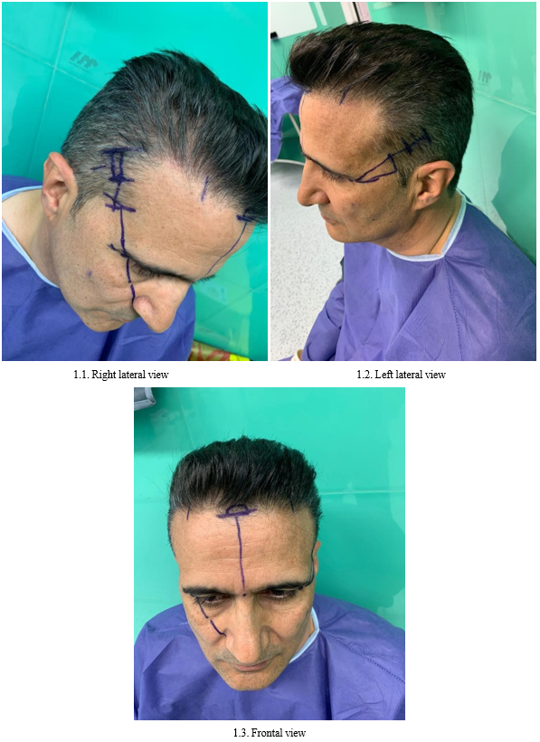
Preoperatively, incision sites were marked in a seated position, including three distinct marks: a central incision and two temporal incisions. The central incision, an elliptical shape (1.00×0.5 cm), started from the nasion, traversed the forehead midline, and terminated just posterior to the hairline. Temporal incisions, 2.00 cm into the hairline, were marked parallel and posterior to the temporal hairline. These sagittal incisions were 1.50 to 2.00 cm long, with landmarks identifying the supraorbital and supraorbital nerves. After general anesthesia, the patient was positioned in reverse Trendelenburg, and a Tumescent solution was injected subperiosteally. This solution aids in pain control and bleeding management. The brow lift involved four stages: incision, dissection, muscle resection, and fixation.
For the elliptical central incision, the surgeon initiates a midline cut through all scalp layers, reaching the subperiosteal plane of the cranium. Subsequently, an elliptical incision (1.50×1.00 cm) in the periosteum creates access ports for endoscopic instruments. Blind dissection is then performed through the midline and paramedian ports, proceeding anteriorly towards a point 1 to 2 cm cephalic to the supraorbital rims and laterally along the lateral orbital rims to the lateral canthus.
Upon completion of periosteum elevation and muscle resection or ablation of the frontalis and corrugators, a curved elevator is introduced via the temporal port to dissect the tissue plane between the temporoparietal fascia (superficial temporal fascia) and the temporalis fascia (deep temporal fascia) overlying the temporalis muscle. Entering this plane is ideally done under direct visualization, releasing the entirety of the superficial temporal space anteriorly, posteriorly, and inferiorly to approximately 1 cm above the zygomatic arch to avoid damaging the facial nerve as it crosses the zygoma.
The sentinel vein, typically located 1 cm lateral to the frontozygomatic suture line, should ideally not be cauterized to minimize the risk of facial nerve injury. Attention must also be paid to this landmark to guide the incision, stopping at this point and continuing down to the inferior orbital rim.
Connection of the lateral and central dissection cavities is achieved by sharply dividing the zone of adhesion at the superior temporal line, often facilitated by passing an elevator from lateral to medial. Similarly, the conjoint tendon is opened above the supraorbital rim and posteriorly until a complete connection is established between the lateral and central dissection pockets. Landmarks are indicated in Figures 1.1 to 1.3.
Figure 1. Landmarks of the operation. 1.1. Right lateral view, 1.2. Left lateral view, and 1.3. Frontal view.
Fixation typically begins at the temporal ports and then at the paramedian ports. A large permanent or semi-permanent suture, such as 4-0 nylon, is employed to secure the temporoparietal fascia to the deep temporalis fascia in a vector following a line from the ala to the lateral canthus. Holes are drilled in the skull through the paramedian incisions to place devices bilaterally. The frontal scalp flap is subsequently lifted off the bone and suspended superiorly onto the device prongs with the aid of an assistant. The surgeon typically opts to place screws at the superior extent of the paramedian incisions, followed by suturing the incisions closed to allow the sutures to pull the scalp back and secure it against the screws. These screws can then be removed in the clinic 40 days post-operatively under local anesthesia. Effective pain management during the initial post-operative period is paramount, with recommendations for ice packs around the eyes, sleeping with a 30-degree head elevation for one week, and the provision of adequate pain medication for the first 24 to 72 hours following surgery.
Variations of the technique
Modifications of this technique include performing the procedure without skin excisions, using suspension sutures attached to the screws, and making incisions or excisions of the corrugator muscle (myotomy or myectomy) in cases of muscle hypertrophy.
Variables
In this study, patient images were assessed to compare the number of wrinkles at two distinct time points: before surgery, and six months post-surgery, all in a rested appearance. These are the usual time appointments in which patients, who underwent brow lift surgery, refer to the hospital to visit the clinicians in Iran. The follow-up examination was conducted at Al-Zahra University Hospital of Isfahan by fellowship students of plastic surgery who had not participated in the surgical procedures.
The same photographic studio took all of the preoperative and postoperative photographs (Canon EOS 500D with Tamron Lens 18- 250; Canon Inc., Ōta, Tokyo). Before performing the imaging procedures, study personnel ensured the neck was free of makeup and jewelry was removed from the treatment area. Digital images of the neck of each subject were obtained at two visits, before treatment, and six months after surgery. For digital imaging, subjects were instructed to adopt neutral, nonsmiling expressions. Subjects were carefully positioned facing the camera for a center view. Two trained raters assessed blinded randomized images of subjects before treatment, and six months posttreatment. After completing the wrinkle assessment, the two raters were unblinded to unify the scores. To assess the clinical outcomes, a combination of two indexes, the Lemperle Wrinkle Assessment Scale [15, 16], and the 5-point Merz Aesthetic Scale [17-19], presented in Table 1, were utilized for this comparison.
Table 1. Combination of Lemperle Wrinkle Assessment Scale and Merz Scale for classification of facial wrinkles.
|
Facial Wrinkle |
Class |
Definition |
|
Lemperle Wrinkle Assessment Scale |
||
|
Horizontal forehead line |
||
|
|
0 |
No wrinkles |
|
|
1 |
Just perceptible wrinkles |
|
|
2 |
Shallow wrinkles |
|
|
3 |
Moderately deep wrinkles |
|
|
4 |
Deep wrinkles, well-dentated edges |
|
|
5 |
Very deep wrinkles, redundant folds |
|
Merz Scale |
||
|
Brow positioning |
||
|
|
0 |
Youthful, refreshed look and high-arch eyebrow |
|
|
1 |
Medium-arch eyebrow; |
|
|
2 |
The slight arch of the eyebrow |
|
|
3 |
Flat arch of the eyebrow, visibility of folds, and tired appearance |
|
|
4 |
Flat eyebrow with barely any arch, marked visibility of folds, and very tired appearance |
|
Glabella lines - at rest |
||
|
|
0 |
No glabellar lines |
|
|
1 |
Mild glabellar lines |
|
|
2 |
Moderate glabellar lines |
|
|
3 |
Severe glabellar lines |
|
Upper cheek fullness – at rest |
||
|
|
0 |
Full upper cheek |
|
|
1 |
Mildly sunken upper cheek |
|
|
2 |
Moderately sunken upper cheek |
|
|
3 |
Severely sunken upper cheek |
|
Combination of Lemperle Wrinkle Assessment Scale and Merz Scale |
||
|
Crew’s feet lines |
||
|
|
0 |
No Wrinkle Crow`s Feet |
|
|
1 |
Mild Wrinkle Lines - Fine Wrinkle |
|
|
2 |
Moderate Wrinkle Lines - Fine to Moderate depth Wrinkles |
|
|
3 |
Severe Wrinkle Lines - Mild Number of Lines |
|
|
4 |
Very Severe Wrinkle Lines -Moderate Number of Lines |
|
|
5 |
Very Deep Wrinkle, Redundant Fold Numerous Lines -Severe Lines |
Additionally, other considered variables included:
1. Demographic features
2. History of any invasive intervention on the face in the past two years (including Botox injection, polydioxanone thread, and soft tissue fillers)
3. The size of scars (the larger one was documented)
4. Satisfaction of patient (assessed by Client Satisfaction Questionnaire (CSQ-8, v. TMS-180S)) [20]
5. Presence or absence of post-surgery complications [21,22] such as:
a. Dimpling at suture
b. Asymmetry
c. Overcorrection
d. Wound dehiscence and necrosis
e. Hematoma
f. Edema or prolonged ecchymosis
g. Obvious scars
h. Complaint of hair loss
i. Transient paresthesia/dysesthesia in the head plus sensory and motor nerve injury in the frontal, supratrochlear, and supraorbital nerves. The patient was asked to describe the sensation of moving and light touch as normal or abnormal.
Results
The patient ages ranged from 35 to 65 years, with an equal distribution of male and female participants. The size of the surgical scars varied between 1 and 2 cm. The most frequently reported complication was paresthesia or dysesthesia in the head, affecting seven out of the eight patients. Despite this, nearly all patients reported high levels of satisfaction with the surgical outcomes.
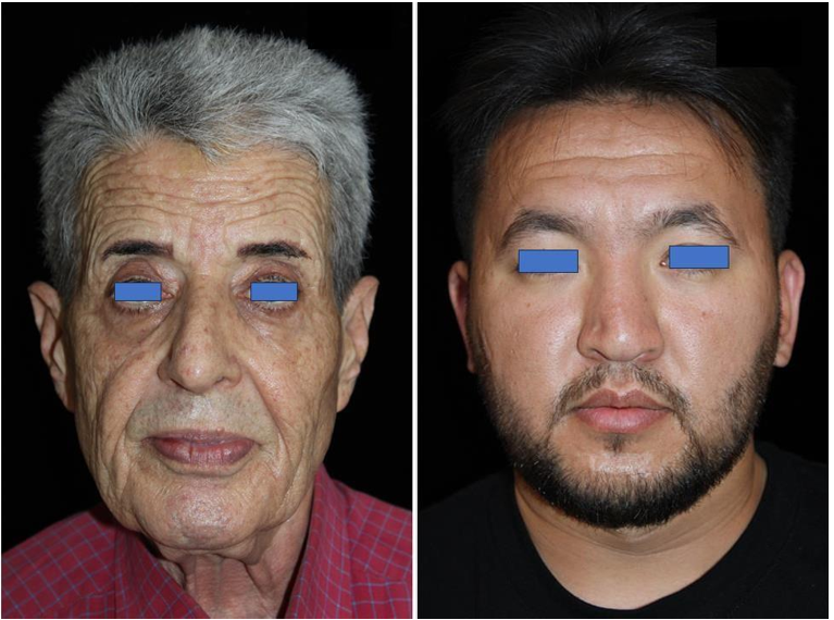
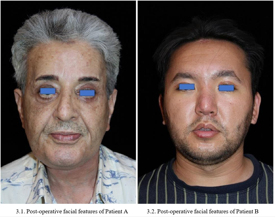
Table 2 outlines the characteristics, aesthetic details, and potential complications of the eight patients who underwent the three-incision endoscopic forehead lift surgery. Also, pre- and post-operative photography of a patient is presented in Figures 2 and 3.
- 2.1. Pre-operative facial features of Patient A 2.2. Pre-operative facial features of Patient B
Figure 2. Pre-operative. 2.1. Pre-operative facial features of Patient A, 2.2. Pre-operative facial features of Patient B.
Figure 3: Post-operative. 3.1. Post-operative facial features of Patient A, 3.2. Post-operative facial features of Patient B.
Table 2: Clinical outcomes of the included patients.
|
Patients’ codes |
Patient One |
Patient Two |
Patient Three |
Patient Four |
Patient Five |
Patient Six |
Patient Seven |
Patient Eight |
||||||||
|
Patient characteristics: |
||||||||||||||||
|
Age |
38 |
35 |
42 |
65 |
42 |
47 |
45 |
40 |
||||||||
|
Gender |
Male |
Female |
Female |
Male |
Male |
Female |
Male |
Female |
||||||||
|
History of any invasive |
Negative |
Negative |
Negative |
Negative |
Negative |
Negative |
Negative |
Negative |
||||||||
|
Outcome Variables: |
||||||||||||||||
|
Primary outcome: |
Pre- |
Post- |
Pre- |
Post- |
Pre- |
Post- |
Pre- |
Post- |
Pre- |
Post- |
Pre- |
Post- |
Pre- |
Post- |
Pre- |
Post- |
|
Lemperle Wrinkle Assessment Scale |
||||||||||||||||
|
Horizontal forehead line |
4 |
1 |
3 |
0 |
5 |
1 |
4 |
1 |
4 |
1 |
4 |
0 |
4 |
1 |
4 |
1 |
|
Merz Scale |
|
|||||||||||||||
|
Brow positioning |
2 |
0 |
2 |
0 |
3 |
0 |
3 |
1 |
2 |
0 |
2 |
0 |
2 |
0 |
2 |
0 |
|
Glabella lines - at rest |
4 |
0 |
2 |
0 |
4 |
0 |
4 |
1 |
4 |
0 |
4 |
0 |
4 |
0 |
4 |
0 |
|
Upper cheek fullness – at |
4 |
1 |
3 |
0 |
3 |
1 |
3 |
1 |
4 |
1 |
4 |
1 |
4 |
1 |
4 |
1 |
|
Combination of Lemperle Wrinkle Assessment Scale and Merz Scale |
||||||||||||||||
|
Crew’s feet lines |
3 |
1 |
3 |
0 |
4 |
0 |
3 |
1 |
3 |
1 |
3 |
1 |
3 |
1 |
3 |
1 |
|
Secondary outcomes: |
||||||||||||||||
|
Size of midline and lateral |
1 |
1.5 |
1 |
2 |
1 |
1 |
1 |
1 |
||||||||
|
Patient’s satisfaction |
30 |
25 |
31 |
28 |
30 |
30 |
31 |
32 |
||||||||
|
Post Operative complications: |
||||||||||||||||
|
Dimpling at suture |
Negative |
Negative |
Negative |
Negative |
Negative |
Negative |
Negative |
Negative |
||||||||
|
Asymmetry |
Negative |
Negative |
Negative |
Negative |
Negative |
Negative |
Negative |
Negative |
||||||||
|
Overcorrection |
Negative |
Negative |
Negative |
Negative |
Negative |
Negative |
Negative |
Negative |
||||||||
|
Wound dehiscence and |
Negative |
Negative |
Negative |
Negative |
Negative |
Negative |
Negative |
Negative |
||||||||
|
Hematoma |
Negative |
Negative |
Negative |
Negative |
Negative |
Negative |
Negative |
Negative |
||||||||
|
Edema or prolonged |
Negative |
Negative |
Negative |
Negative |
Negative |
Negative |
Negative |
Negative |
||||||||
|
Obvious scars |
Negative |
Negative |
Negative |
Negative |
Negative |
Negative |
Negative |
Negative |
||||||||
|
Complaint of hair loss |
Negative |
Negative |
Negative |
Negative |
Negative |
Negative |
Negative |
Negative |
||||||||
|
Transient paresthesia/ |
Positive |
Positive |
Negative |
Positive |
Positive |
Positive |
Positive |
Positive |
||||||||
Discussion
While non-surgical methods are temporary, require less technical expertise, and allow for easier correction in case of complications, surgical approaches—particularly the endoscopic forehead lift— are favored by individuals more accepting of the potential for scarring. This includes patients such as males or those with high hairlines, for whom minor scars are less noticeable [4]. However, Perenack [11] argued that the endoscopic forehead lift may benefit individuals with a normal to short hairline.
According to him, the ideal candidates for this procedure are those who also exhibit brow ptosis or have excess forehead or temporal skin, as these conditions can be effectively addressed through surgery. In addition, Isse [12] emphasized that the technique is especially advantageous for patients with thin or balding hair, as the minimal incision scars are easily hidden, making it a more appealing option for this demographic. Collectively, these perspectives suggest that while the endoscopic forehead lift is versatile, patient selection based on hairline, skin condition, and brow positioning is crucial for optimal results.
Isse [12] observed that individuals with tight forehead skin, commonly seen in patients of Latina, Asian, and American Indian descent, are generally not ideal candidates for the endoscopic forehead lift procedure. This is due to the reduced skin elasticity in these populations, which can limit the effectiveness of the technique and potentially lead to suboptimal outcomes. In contrast, in the present study, none of the participants fell into these demographic categories, as such cases are extremely rare within the Iranian population. This demographic difference highlights the importance of tailoring surgical techniques to the specific characteristics of the patient population to ensure the best possible outcomes, as certain ethnicities may present unique anatomical challenges that could influence the success of the procedure.
The endoscopic technique has become a common approach in forehead rejuvenation, though it requires advanced training and specialized equipment to perform effectively [12]. In some instances, the results may be moderate or may not endure over time [4]. However, Behmand et al. [9] counter this by emphasizing that, when performed correctly, endoscopic forehead rejuvenation yields stable and lasting outcomes, with patients maintaining improvements for many years post-surgery. Furthermore, de la Fuente et al. [23] introduced another crucial factor that can influence the success of the procedure: the proximity of the brow fixation point. According to their findings, the closer the fixation point is to the brow, the greater the degree of tissue advancement, leading to more significant and durable postoperative results. This highlights the importance of precise technique and planning in ensuring both the immediate and long-term effectiveness of the surgery.
Consistent with the findings of the current study, both De Cordier et al. [24] and Wang et al. [25] reported that the endoscopic forehead lift effectively reduces transverse forehead wrinkles and alleviates glabellar frown lines while elevating the eyebrows. These improvements not only enhance the aesthetic appearance but also contribute to a more youthful and refreshed look, diminishing the tired or angry expressions that can be associated with aging. By opening up the area around the eyes, the procedure significantly enhances facial expression and overall appearance, reinforcing the positive outcomes noted in our research. This highlights the endoscopic forehead lift as a valuable option for individuals seeking to rejuvenate their facial aesthetics.
While Wang et al. [25] utilized the FACE-Q scales to assess patient outcomes, our study employed the Client Satisfaction Questionnaire (CSQ-8, version TMS-180S) to evaluate patient satisfaction. Despite the use of different instruments, both studies demonstrated consistently high levels of satisfaction among patients. This suggests that, regardless of the specific tool used for measurement, patients generally express favorable opinions regarding the results of the endoscopic forehead lift, reinforcing the procedure's effectiveness in meeting aesthetic and clinical expectations. The similarity in findings across different assessment methods further validates the positive impact of this surgical intervention.
In line with our findings, Behmand et al. [9], Cho et al. [13], and de la Fuente et al. [23] also reported that the most common complication associated with the endoscopic forehead lift was some degree of transient or persistent paresthesia. Isse [12] further noted that the typically temporary nature of numbness is an advantage of this procedure, as permanent numbness is rare. In addition to paresthesia, these studies highlighted other complications such as dynamic forehead irregularities, asymmetry in movement [9,13], itching sensations [9], and alopecia [13]. However, in our study, none of the patients experienced alopecia, and no significant forehead irregularities or other complications were observed. These findings suggest that while some complications are common in the literature, the outcomes may vary depending on surgical technique, patient characteristics, and postoperative care.
The limitations of this study include its retrospective design and the absence of a direct comparison with other surgical or non-surgical techniques. The lack of a control group prevents a more comprehensive evaluation of the relative effectiveness and complication rates of the endoscopic forehead lift compared to alternative methods. Additionally, the small sample size restricts the generalizability of the findings to a broader population. To address these limitations, future research should focus on conducting case- control studies and randomized clinical trials with larger sample sizes. A longer follow-up period would also provide more insight into the long-term outcomes and stability of results, especially considering that previous studies, such as those by Behmand et al. [9] and de la Fuente et al. [23], suggest the potential for stable results over many years. Incorporating comparisons between different techniques and evaluating factors such as patient satisfaction, complication rates, and postoperative outcomes would offer a more robust understanding of the effectiveness of the endoscopic forehead lift in various patient populations.
Conclusion
The study assessed eight patients who underwent the three-incision endoscopic forehead lift, with ages ranging from 35 to 65 years and an equal number of males and females. Surgical scars varied from 1 to 2 cm, and the most frequently reported complication was paresthesia, noted in seven patients. Notably, despite the presence of this complication, nearly all patients reported high levels of satisfaction with their overall outcomes, reflecting the procedure's success in enhancing facial aesthetics.
Funding Source: This research project was partially funded by Isfahan University of Medical Sciences.
Declaration of AI and AI-assisted technologies in the writing process?
There is nothing to disclose.
Compliance with ethical approval
Conflict of Interest Statement: The authors declare no conflicts of interest to disclose.
Statement of human and animal rights, or ethical approval: This Research study is approved by the Ethical Committee of the Isfahan University of Medical Sciences (IR.MUI.MED.REC.1401.426).
Informed consent: Informed consent was taken from all included participants in their native language, Persian.
References
- Finn CJ, Cox SE, Earl ML (2003) Social implications of hyper functional facial lines. Dermatol Surg. 29(5): 450-455.
- Caruthers J, Glogau RG, Blitzer A. (2008) Advances in facial rejuvenation: botulinum toxin type A, hyaluronic acid dermal fillers, and combination therapies-consensus recommendations. Plast Reconstr Surg. 121(5 Suppl): S5-S30.
- Ko HJ, Choi JY, Moon HJ, Lee JW, et al. (2016) Multi- polydioxanone (PDO) scaffold for forehead wrinkle correction: a pilot study. J Cosmet Laser Ther. 18(7): 405-408.
- Karimi N, Kashkouli MB, Sianati H, et al. (2020) Techniques of eyebrow lifting: a narrative review. J Ophthalmic Vis Res 15(2): 218-235.
- Chatrath V, Banerjee PS, Goodman GJ, Rahman E (2019) Soft- tissue Filler-associated Blindness: a systematic review of case reports and case series. Plast Reconstr Surg Glob Open. 7(4): e2173.
- Dayan SH, Arkins JP, Mathison CC (2011) Management of Impending Necrosis Associated with soft tissue filler injections.J. Drugs Dermatol. 10(9): 1007-1012.
- Lewis MB (2018) The interactions between botulinum-toxin- based facial treatments and embodied emotions. Sci Rep. 8(1): 14720.
- Lafaille P, Benedetto A (2010) Fillers: contraindications, side effects and precautions. J Cutan Aesthet Surg. 3(1): 16-9.
- Behmand RA, Guyuron B (2006) Endoscopic forehead rejuvenation: II. Long-term results. Plast Reconstr Surg. 117: 1137-1143.
- Stanek JJ, Berry MG (2014) Endoscopic-assisted brow lift: revisions and complications in 810 consecutive cases. J Plast Reconstr Aesthet Surg. 67(7): 998-1000.
- Perenack JD (2016) The endoscopic brow lift. Atlas Oral Maxillofac Surg Clin North Am. 24(2): 165-173.
- Isse N.G (2020) Endoscopic facial rejuvenation: endoforehead, the functional lift. case reports. Aesth Plast Surg. 44: 1162-1170.
- Cho MJ, Carboy JA, Rohrich RJ (2018) Complications in brow lifts: a systemic review of surgical and nonsurgical brow rejuvenation. Plast Reconstr surg Open. 6(10): e1943.
- Graham DW, Heller J, Kirkjian TJ, Schaub TS, Rohrich RJ (2011) Brow lift in facial rejuvenation: a systematic literature review of open versus endoscopic techniques. Plast Reconstr Surg. 128: 335e-341e.
- Lemperle G, Holmes RE, Cohen SR, Lemperle SM (2001) A classification of facial wrinkles. Plast Reconstr Surg. 108(6): 1735-50.
- Flynn TC, Carruthers A, Carruthers J, Geister TL, Görtelmeyer R, et al. (2012) Validated assessment scales for the upper face. Dermatol Surg. 38(2 Spec No.): 309-19.
- Stella E, Di Petrillo A (2014) Standard Evaluation of the Patient: The Merz Scale. In Injections in Aesthetic Medicine, ed 1. Springer, Milano, 33-50.
- Muti GF, Basso M (2017) Treatment of lateral periorbital lines with Different Dilutions of IncobotulinumtoxinA. J Clin Aesthet Dermatol. 10(9): 27–29.
- Carruthers A, Carruthers J, Hardas B, Kaur M, Goertelmeyer R, et al. (2008) A validated brow positioning grading scale. Dermatol Surg. 34(Suppl 2): S150-4.
- Larsen DL, Attkisson CC, Hargreaves WA, Nguyen TD (1979) Assessment of client/patient satisfaction: development of a general scale. Evaluation and Program Planning. 2(3): 197-207.
- Raggio BS, Winters R (2023) Endoscopic Brow Lift. In: StatPearls [Internet]. Treasure Island (FL): StatPearls Publishing.
- Hu X, Ma H, Xue Z, Qi H, Chen B (2017) Endoscopic facelift of the frontal and temporal areas in multiple planes. Singapore Med J. 58(2): 107-110.
- de la Fuente A, Santamaría AB (2002) Endoscopic forehead lift: is it effective?, Aesthet Surg J. 22(2): 113–120.
- De Cordier BC, de la Torre JI, Al-Hakeem MS, Rosenberg LZ, Gardner PM, et al. (2002) Endoscopic forehead lift review of technique, cases, and complications. Plast Reconstr Surg. 110(6): 1558-1568.
- Wang G, Tao L, Xie H (2020) A novel endoscopic forehead lift technique for patients with upper facial wrinkles: morphometric evaluation and patient-reported outcome using FACE-Q Scales Facial Plast Surg. 36(3): 226-234.


