


Rajaa El azzouzi1,2*, Othmane Bouanani1,2, Amine Oussalem1,2, Malik Boulaadas1,2
1Department of Maxillofacial Surgery and stomatology, Hospital of Specialities Rabat, Morocco.
2Faculty of Medicine and Pharmacy of Rabat. Mohammed V University in Rabat, Rabat, Morocco.
*Corresponding Author: Rajaa El azzouzi, Department of Maxillofacial Surgery and stomatology, Hospital of Specialities RABAT ; CHU Ibn Sina, Av. Abderrahim Bouabid, Rabat-Morocco.
Abstract
Introduction
Pindborg tumor, or calcifying epithelial odontogenic tumor (CEOT), is a rare benign tumor. Two clinical-topographic variants are known of this tumor: Intraosseous (central) or Extraosseous (peripheral. The intraosseous tumors have a predilection for the mandible. The radiological diagnosis is usually guided by the presence of calcification images confirmed by the histopathological study.
Case report
A 25-year-old man presented with a Pindborg tumor of the mandible with an intraosseous variant; this case illustrates the difficulty of diagnostic orientation when the radiological appearance is not very specific. The anatomopathological examination concluded with the diagnosis.
Discussion
Pindborg tumor is a rare tumor with a preferential posterior mandibular location. Its calcified character on radiographs usually characterizes it. There are two histological forms: A squamous variant with a favorable evolution and a clear cell variant with a more uncertain prognosis.
Conclusion
The available evidence suggests that this tumor is somewhat less aggressive than ameloblastoma but that any treatment not including excision or fulguration of the rim of the bone surrounding the lesion is likely to allow a recurrence. No instance of metastasis from this neoplasm has been reported.
Keywords: Calcifying epithelial, Odontogenic tumor, CEOT, Pindborg.
Introduction
Calcifying epithelial odontogenic tumor, or Pindborg tumor, is a rare benign tumor. Two clinical-topographic variants are known of this tumor: Intraosseous (central) or Extraosseous (peripheral). The intraosseous tumors have a predilection for the mandible. Imagery shows various forms that lead to the diagnosis, which is only confirmed by histopathology study; we report a slow-growing Pindborg tumor in a young patient revealed by infection complication.
Case report
We report a case of 25 years old man with no medical history who consulted for a painful left jugal tumefaction evolving for 12 months associated lately with an inflammatory sign and dental mobility. (Figure 1) The clinical examination found a facial asymmetry with a painful left jugal mass, poorly limited with bony consistency measuring approximately 65 mm of the long axis, with no inflammatory signs of the skin associated with labio-mental anesthesia. Intraoral examination revealed a gingival expression of the mass filling the lower vestibule, ranging from the 44th to 47th tooth, causing their mobility. An Orthopantomogram showed a radiotransparent lesion with a “soap bubble” texture extending from the 44th to the mandibular ramus, including the angle with an impacted tooth and root resorption of the 45, 46, and 47th teeth. (Figure 2)
An intra-oral biopsy was performed under local anesthesia, for which the histopathological examination revealed characteristic features of the calcifying epithelial odontogenic tumor (Pindborg tumor). Accordingly, surgery was planned under general anesthesia to resect the lesion with a minimum of 1 cm margin, using a vestibular approach (Figure 3,4).
No reconstruction was needed. The postoperative course was straightforward, with no recurrence at 6 months follow-up.
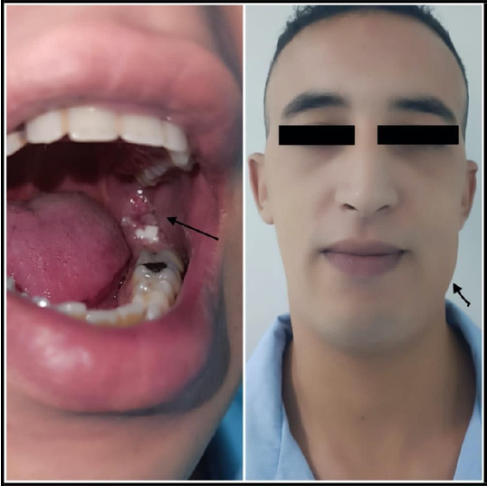
Figure 1: Pre-operative images showing the mandibular tumor.

Figure 2: OPT image showing the extension of the Pindborg tumor
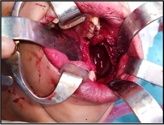
Figure 3: Per-operative image after the excision of the tumor
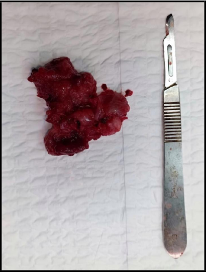
Figure 4 : Macroscopic appearance of Pindborg tumor
Discussion
A calcifying epithelial odontogenic tumor (CEOT) is a rare benign odontogenic tumor called the ‘‘Pindborg tumor’’, as Pindborg first described it in 1955. The origin of this rare, aggressive, and locally invasive tumor remains unknown.
Pindborg first described the calcified odontogenic epithelial tumor in 1955; it represents less than 1 % of odontogenic tumors [1,2,3]. CEOTs occur over a broad age range; however, they are slightly more common between 3rd and 6th decades of life [4]. There is no gender predilection.
The origin of this tumor remains debated. Pindborg [3] and Ficarra [5] have proposed the reduced enamel epithelium as a possible origin. This hypothesis is related to the frequent association of this tumor with an unerupted or embedded tooth, as represented in our case.
Pindborg tumor is a rare and benign odontogenic neoplasm that affects the jaw. It can be divided into 2 clinical-topographic variants: Intraosseous (central) or Extraosseous (peripheral), with an incidence of 94 % and 6 %, respectively. Intraosseous tumors have a predilection for the mandible, and most tumors arise in the molar and premolar regions of the mandible. When present, extraosseous tumors are often located in the anterior jaw and involve the gingiva.
The main form is the most common, usually manifesting as an indolent, slow-growing swelling; it grows by infiltration, producing cortical expansion, tooth movement, and root resorption [6]. However, maxillary localizations can be the source of pain, nasal obstruction, and epistaxis. In the reported case, the incidental finding and the discretion of the symptomatology reflect the low aggressive nature of the lesion, and the infection was the reason for consultation and the alarm signal.
The radiographic appearance of CEOT is variable and depends on the stage of development. It includes unilocular pericoronal radiolucency and multilocular radiolucent images associated with tiny pictures of calcifications that increase in size with time associated with an unerupted or embedded tooth [6].
The absence of a radiopaque image poses the problem of differential diagnosis, particularly with other benign intraosseous tumors such as ameloblastoma, odontogenic keratocyst, or giant cell granuloma. As represented in our case, there a biopsy was performed.
Histopathology is the most variable aspect of this tumor; There are sheets of polyhedral epithelial cells with eosinophilic cytoplasm, showing a moderate degree of pleomorphism but only rare typical mitoses. A cellular hyalinised stromal bridge, interspersed with foci of an amyloid-like substance, may be seen [4,7,8]. Other variants, such as a noncalcifying CEOT with Langerhans cells, which is reportedly more aggressive, CEOT with cementum and bone-like materials, and a clear cell variant of CEOT, have also been described [9,10].
CEOT has to be treated surgically but depending upon the lesion, simple curettage, enucleation, marginal resection with tumor-free average margins, or hemimandibulectomy, and hemimaxillectomy is performed. However, CEOT is considered less aggressive clinically than typical infiltrating ameloblastoma and is deemed to have a rate of recurrence much lower than that of ameloblastoma. Hence Such cases need to have a long-term follow-up to rule out recurrence; It seems that those treated with enucleation and curettage procedures show a recurrence rate ranging from 15 % to 30 % after just 2e4 years.
Conclusion
Pindborg tumor is an uncommon odontogenic tumor with a characteristic variation in its histopathologic, clinical, and radiologic presentation. Thereby posing a diagnosis is challenging. Being a locally aggressive tumor, resection with adequate average margins is mandatory when treating large lesions to prevent a recurrence. Proper reconstruction of the bony defect and subsequent follow-ups are required to decrease the morbidity associated with large tumors.
Informed consent: Free and informed consent has been given by the patient.
Conflicts of interest: The authors declare no conflicts of interest.
References
- Franklin CD, Pindborg JJ (1976) The calcifying epithelial odontogenic tumor. A review and analysis of 113 cases. Oral Surg Oral Med Oral Pathol. 42(6): 753-65.
- Basu MK, Matthews JB, Sear AJ, Browne RM (1984) Calcifying epithelial odontogenic tumor: a case showing features malignancy. J Oral Pathol. 13(3): 310-9.
- Pindborg JJ (1958) A calcifying epithelial odontogenic tumor. Cancer. 11(4): 838-43.
- Rajendran R. Cysts and tumors of odontogenic origin. In: Rajendran R, Sivapathasundharam B, editors. Shafer’s Textbook of Oral Pathology. 6th ed. New Delhi: Elsevier; 2009. p. 278-81.
- Ficarra G, Hansen LS, Stiesmeyer EH (1987) Intramural calcifiying epithelial odontogenic tumor. Int J Oral Maxillofac Surg. 16(2): 217-21.
- Kaplan I, Buchner L, Calderon S, Kafe I (2001) Radiological and clinical features of calcifying epithelial odontogenic tumour. Dentomaxillofac Radiol. 30(1): 22-8.
- Reichart PA, Philipsen HP (2004) Calcifying epithelial odontogenic tumors. In: Reichart PA, Philipsen HP, editors. Odontogenic Tumors and Allied Lesions. London: Quintessence Publishing. 93-104.
- Waldron CA. Odontogenic cysts and tumors. In: Neville BW, Damm DD, Allen CM, Bouquot JE, editors. Oral and maxillofacial pathology. 3rd ed. Philadelphia: W.B. Saunder; 2008. p. 623-5.
- Sahni P, Nayak MT, Singhvi A, Sharma J (2012) Clear cell calcifying epithelial odontogenic (Pindborg) tumor involving the maxillary sinus: A case report and review of literature. J Oral Maxillofac Pathol. 16(3): 454-9.
- Kamath G, Abraham R (2012) Recurrent CEOT of the maxilla. Dent Res J. 9(2): 233-6.