


Ray David Molina Linares1*, A.S. Mary Gabriela Teran Barreto1, Eduardo Ignacio Madrid Vasquez2, Rafael Jose Santiago Pena3
1Pediatrician and Child Care Specialist, University of Los Andes, Venezuela.
2Surgeon, University of Los Andes. Venezuela.
3Pediatric Gastroenterologist. University of Los Andes, Venezuela.
*Corresponding Author: Ray David Molina Linares, Pediatrician and Child Care Specialist, University of Los Andes, Venezuela.
Abstract
Introduction: The term leishmaniasis is the name given to a group of diseases caused by the protozoan of the genus Leishmania, transmitted to humans by an infected sand fly vector. Visceral leishmaniasis (VL) is the most serious clinical presentation. It is characterized by fever, splenomegaly, hepatomegaly and alterations such as anemia, leukopenia, hypoalbuminemia. Clinical case: We present a 3-year-old female preschooler, who presented with abdominal distension and long-standing bloody stools. There was no visceromegaly during the course of the disease. The studies requested and the epidemiological history raised the suspicion of atypical VL, confirming the disease with a bone marrow aspirate. Treatment with Glucantime was completed for 14 days and Amphotericin B was continued. Conclusion: Inexperience in diagnosis, clinical suspicion and particularly, its epidemiology, can lead to a delay in the timely management of the disease, leading to the risk of life for the patient.
Keywords: Visceral leishmaniasis, hepato-splenomegaly.
Introduction
The term leishmaniasis is the name given to a group of diseases caused by the intracellular protozoan of the genus Leishmania, transmitted to humans by the bite of an infected sand fly insect vector [1]. It includes a clinical spectrum that encompasses cutaneous, mucosal and visceral lesions [2]. Leishmaniasis is endemic in 88 countries around the world, especially in countries with tropical and temperate zones [3]. In the region of the Americas, leishmaniasis continues to be a public health problem due to its wide magnitude, geographic distribution, morbidity and mortality [4].
Visceral leishmaniasis (VL) is the most serious clinical presentation of the disease, with a fatality rate of 90% without treatment [3]. The importance of this disease in the pediatric age lies in the high vulnerability that children have to infection, the difficulties and errors in diagnosis, which results in greater injury and tissue damage [1]. In our country, the first case of VL was described in 1941, where the chain of transmission was studied (sandflies as vector, canines as reservoirs and the human population as those exposed to risk), with special attention being given to the western focus, in the states of Lara, Trujillo and Falcón [5].
Subsequently, studies have been carried out with special attention to specific epidemiological foci, for example, in the state of Falcón, where there was a strong tendency towards under-reporting of leishmaniasis cases over a prolonged period of time [6]. Likewise, in the state of Nueva Esparta, for the year 2006, a significant increase in visceral leishmaniasis was observed in pediatric patients, with a trend that reaches its maximum in the group of 1-4 years of age, being related to risk factors such as sanitary deficiency. , malnutrition and inadequate practices in relation to the environment [7].
In relation to the state of Trujillo, a trend towards poor notification of leishmaniasis cases was evident. This state is considered the third with the highest incidence of visceral leishmaniasis, especially in pediatric patients, where the presentation of the disease is of high incidence in the age group between 0-5 years, with a predominance of the male sex [8]. It is vitally important to recognize that the studies carried out in our country on leishmaniasis in pediatrics are scarce and those carried out are more than 5 years old, so the impact in Venezuela and the Americas, on the knowledge of this disease, should raise awareness. local researchers to expand knowledge of the natural history of this pathology, which will contribute to developing prevention and control strategies [9].
Unlike the cutaneous form, the clinical picture of VL is very similar throughout the world. The average incubation period is three to eight months [1]. VL is clinically characterized by fever, anorexia, weight loss, splenomegaly, hepatomegaly and laboratory abnormalities, such as anemia, leukopenia, thrombocytopenia, hypoalbuminemia and hypergammaglobulinemia [3]. VL should be suspected in all pediatric patients with a fever of unknown origin [10]. Multiple factors favor the re-emergence of this pathology worldwide: climate changes and deforestation, causing greater exposure to vectors, disorganized migrations, urbanizations lacking environmental sanitation, and regional trends toward tropicalization [11].
A clinical case is presented that generated conflict in the diagnosis and motivated specialists, residents and students to review the clinical manifestations of VL in our environment, as well as the atypical forms of the disease, timely treatment, adverse reactions to said treatment and clinical resolution favorable.
Clinical Case
A 3-year-old female preschool patient, native and from Caja Seca, Zulia state, who is taken to the pediatric emergency of the hospital by her mother, reporting the onset of a current illness characterized by liquid stools with mucus and blood of 1 month of evolution, with number 6 – 7 bowel movements per day, accompanied by an unquantified increase in body temperature and abdominal pain of mild to moderate intensity, diffuse, without irradiation, going to the local doctor (Caja Seca) on multiple occasions, who indicated metronidazole, albendazole and secnidazole, obtaining relative improvement. However, in view of the persistence of the clinical picture, she went and it was decided to admit her. The mother denies any significant medical history. She refers to her epidemiological history as living in an improvised house, built with bamboo cane, a tin roof, with the use of a latrine, with two rooms for 6 people. They have domestic animals, two dogs, and fattening animals such as pigs and cows, approximately 15 animals. According to maternal reference, more than 5 dog deaths due to probable bloody stools have been reported in the area.
On physical examination, respiratory rate: 23 rpm, heart rate: 132 bpm, Temperature: 37.4 °C, blood pressure: 112/60 mmHg, Weight:
10.5 Kg, Height: 86 cm, patient in regular clinical condition, afebrile, eupneic, tachycardic, tolerating ambient oxygen, with signs of mild dehydration, dry oral mucosa, with stringy saliva. Thin hair with flag sign. Rhythmic heart sounds without murmurs, audible vesicular murmur in both lung fields without aggregates, without signs of respiratory distress. Abdomen slightly distended, fluid sounds present, painful on superficial palpation, no visceromegaly palpable.
Limbs symmetrical, mobile, without presence of edema. Neurological: alert, active, without motor or sensory deficit, cooperative with the physical examination.
Hemoglobin 5.6 gr/dl; white blood cells 17,900 /mm3; neutrophils 44%, lymphocytes 56%; platelets 126,000 /mm3. It was decided to corroborate hemoglobin levels, so a blood count was requested, which reported: Hemoglobin 3.3 gr/dl; hematocrit 11.9%; white blood cells 13,100 /mm3, neutrophils 57%; lymphocytes 26%; eosinophils 11%; crooks 4%; platelets 335,000/mm3, increased in cresil. Predominance of microcytes and macrocytes. As indicated, globular concentrate on 2 occasions. An abdominal ultrasound is requested and reports: liver, spleen and pancreas in normal shape and size. No presence of ascites or intra-abdominal masses (No obvious pathological findings). An evaluation is requested by the Hematology service who, in view of severe anemia, the patient's origin, and the presence of atypical microorganisms in blood biometry, requests to rule out VL, requesting a formolgelation test, bone marrow aspirate, and indirect markers that include lactate dehydrogenase. , total and fractionated bilirubin, uric acid, alkaline phosphatase and serum electrolytes.
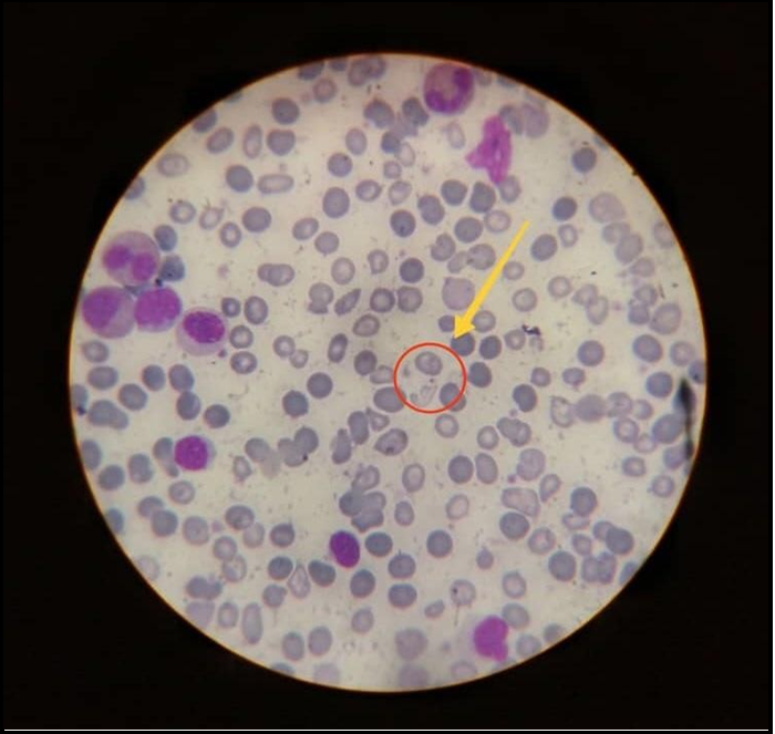
Subsequently, due to a positive formolgelation test, an evaluation was requested by the pediatric infectious disease service, who decided to wait for a bone marrow aspirate to start treatment with antimonials. However, they decided to request febrile antigens, rule out malaria and Chagas disease due to the patient's origin. , request HIV and VDRL, BK of gastric contents, all of which were performed and reported as negative. The patient is taken to the operating room to perform a bone marrow aspirate under sedation, where a week later, a report is received on the smear that is positive for Leishmania, with moderate amounts of amastigotes observed (Figure 1). Treatment was started with N-methyl-glucamine antimoniate (Glucantime) at a dose of 20 mg/kg/day deep intramuscularly every other day for 20 doses. However, in view of presenting signs of cardiotoxicity of Glucantime upon administration of the 14th dose of the same, it was decided to change the treatment to liposomal Amphotericin B at a dose of 4 mg/kg/day for the first 5 days. Her evolution was favorable, and she was discharged with monthly follow-up by the Pediatric Infectious Diseases service.
Discussion
Visceral leishmaniasis in pediatrics is typically characterized by a pattern of intermittent fever, abdominal distention, hemorrhages, weight loss, and cutaneous-mucosal pallor, with physical examination findings of hepatosplenomegaly and generalized lymphadenopathy, as is the case reported by Álvarez et al. [1]. However, in the case presented, there were no abdominal megalias during the course of the disease, giving it the name atypical visceral leishmaniasis.
Although it is believed that VL is still considered a tropical disease, its endemicity should be considered, as reported by Gomila et al [11]. Since Venezuela is an endemic country for Leishmaniasis, in any patient with suspected symptoms of intermittent fever, hemorrhages and abdominal distention, VL must be ruled out, as occurred in the aforementioned case.
In most patients, analytical data guide the diagnosis; however, in the visceral form, the sample for direct examination can be obtained by bone marrow aspirate, presenting 97% sensitivity and specificity [4]. In the case described, the diagnosis of the disease was suspected and finally confirmed with the positivity of the formol gelation test and the bone marrow aspirate, considering the latter as the most reliable and safest [10].
The treatment used in Venezuela is standardized to the therapeutic standards suggested by the Pan American Health Organization (PAHO) [4]. The drugs used are pentavalent antimonials and amphotericin B, although the latter has been conceived as first-line in the European continent, as reported by Gomila et al [11]. In the patient presented, Glucantime was used, resulting in a cardiotoxic and potentially serious reaction, which could be prevented under monitoring and control. Context that in multiple research articles describes the main mechanism of cardiotoxicity of antimonials as the prolongation of the QT interval and the alteration of the morphology of the T wave in the electrocardiographic recording, with the potential development of fatal arrhythmias [12]. Likewise, she received liposomal amphotericin B, with an excellent therapeutic, clinical and hematological response. Drug described as an effective, safe treatment with no relapses in 12 months of follow-up for VL [3]. Liposomal amphotericin B is described as the treatment with the least toxicity compared to amphotericin B deoxycholate and pentavalent antimonials [13].
In pediatrics, leishmaniasis, and especially the visceral form, should be considered as a differential diagnosis, in multiple pathologies that present with similar symptoms, whether or not they have hepato- splenomegaly. Inexperience in diagnosis, clinical suspicion and particularly, its epidemiology, can lead to a delay in the timely management of the disease, leading to risk of life for the patient.
References
- Álvarez Peñalosa E, Paláu-Castaño J. Infections in pediatrics. Prevention, diagnosis and treatment. Second edition. Inter- American Edit. Bogotá 2003, 785p.
- Rivero-Rodríguez ME, Pérez-Doria A, Bejarano-Martínez EE (2020) Visceral leishmaniasis in children and canine populations in the urban area of the municipality of Ovejas, Colombia. Science and innovation in health. 95: 357-358.
- Mañes Jiménez Y, Pedrón Marzal GM (2021) Analysis of 37 cases of Leishmaniasis in children, diagnosed in a region of Valencia, Spain. Rev Pediatr Aten Primary. 23: 33-41.
- Pan American Health Organization (2022) Guideline for the treatment of leishmaniasis in the Americas. Second edition. Washington, D.C. 24p.
- Traviezo L (2012) Diversity of sandflies in an endemic area of American visceral leishmaniasis in Venezuela. Rev Peru Med Esp Public Health. 29(4): 503-508.
- Vargas E, Yépez J (2004) Epidemiological aspects of visceral leishmaniasis in Venezuela, with special attention to the Falcón State. Bol Mal Salud Amb. 44(1): 9-19.
- Moreno M, Ortega M, Belizario D, Galindo W, Zerpa O (2016) Epidemiology of Visceral Leishmaniasis in the state of Nueva Esparta, Venezuela. Period 1995-2015. Dermatol Venezuela. 54(1): 17-23.
- Vásquez L, Vásquez L, Oviedo M, Sandoval C, Méndez Y, et al. (2010) Clinical and epidemiological profile of American visceral leishmaniasis in the state of Trujillo, Venezuela (1975-2007). Bol Mal Salud Amb. 50(2): 233-242.
- Isaza-Jaimes A, Rodríguez JE, Chacón G, Bravo A, Silva C (2018) A vision about American Leishmaniasis and its epidemiological behavior. 37(3): 190-196.
- Sagrós A, Subirón R, Guerrero C, De Arriba A, Bustillos M (2018) Pediatric visceral leishmaniasis. About a case. Bol Pediatr Arag Rioj Sor. 48: 85-87.
- Gomila H A, Vanzo C, Garnero A, Peruzzo L, Badalotti M (2017) Visceral leishmaniasis. Pediatric clinical case. Arch Argent Pediatr. 115(4): 251-254.
- Arcos L, Rincón C, Vanegas D, Medina R (2081) Electrical storm and torsade de pointes associated with treatment with antimonials in a patient with cutaneous leishmaniasis. Rev Colomb Cardiol. 25(4): 279e1-279e5.
- Vélez I, Jimenez A, Cachelix C, Vásquez D, López L (2012) Therapeutics of American Leishmaniasis. New recommendations. Dermatol Venezuela. 50(1): 15-18.