


David Cajthaml2, Alison Ingraldi2, Josh Sandberg1,2, Aaron J. Tabor1,2*
1Department of Medical and Technical Sciences, Medtech Incubator, Scottsdale, AZ 85260, USA
2Department of Research & Clinical Sciences, Axolotl Biologix, Scottsdale, AZ 85260, USA
*Corresponding Author: Aaron J. Tabor, Department of Medical and Technical Sciences, Medtech Incubator, Scottsdale, AZ 85260, USA and Department of Research & Clinical Sciences, Axolotl Biologix, Scottsdale, AZ 85260, USA
Abstract
Background: Diabetic foot ulcers (DFUs) remain difficult to heal despite comprehensive standard care, and advanced wound-care strategies are frequently evaluated as adjuncts when wounds fail to progress. AXOLCAMP is an ongoing multicenter, open-label, randomized controlled trial evaluating a product specific dehydrated human amnion membrane (dhAM), Axolotl DualGraft™, plus standard of care (SOC) versus SOC alone in nonhealing DFUs.
Methods: Adults with type 1 or type 2 diabetes and an eligible nonhealing DFU are randomized to dhAM plus SOC or SOC alone. The protocol targets 100 evaluable participants (50 per group), with recruitment of approximately 112–115 participants to account for anticipated attrition. The primary endpoint is the proportion of target ulcers achieving complete wound closure within 12 weeks. Secondary endpoints include time to closure, percent area reduction (PAR), adverse events, pain, and wound-related quality of life. This manuscript reports a mid-study analysis, using data provided on 23 September 2026. These data sets do not include the complete randomized schedule which has yet to be fully enrolled as the study is still ongoing.
Results: At the supplied operational snapshot, 171 participants had been screened, 86 had been randomized, 46 had completed the study, and 25 remained ongoing. Apparent complete 12-week wound closure from completed subjects (protocol modification without confirmation) for the SOC and SOC + Treatment groups was observed in 3/19 (15.8%) and 5/27 (18.5%), respectively. Mean PAR at 12 weeks was 19.16% versus 41.07%, respectively. Safety outcomes were similar across both groups (SOC = 31.8%, SOC + Treatment = 33.3%). Pain, Enjoyment of Life, and General Activity (PEG) scores at week 12 were significantly lower in the dhAM + SOC group than in the SOC-alone group (1.31± 2.11 vs. 2.86 ± 2.97; p = 0.044). Between-group change from baseline did not differ (p = 0.774)
Conclusions: This mid-study analysis provides an early randomized comparison of dhAM plus SOC versus SOC alone in nonhealing DFUs. Preliminary findings suggest an increase in PAR with adjunctive dhAM, while its effect on complete wound closure remains uncertain. Because enrollment and follow-up remain ongoing, these findings should be interpreted as exploratory and do not replace the prespecified final analysis of the completed trial.
Keywords: CAMPs; diabetic foot ulcer; dehydrated human amnion membrane; amniotic membrane; randomized controlled trial; wound closure; percent area reduction; standard of care; AXOLCAMP
Introduction
Diabetic foot ulcers and their infectious complications are major contributors to diabetes-related lower-extremity morbidity, recurrent ulceration, and amputation [1–3]. In one large prospective study of infected diabetic foot ulcers (DFUs), fewer than 50% had healed within one year, and 10% of those that healed later recurred [2]. Beyond failure to heal, the long-term burden of DFUs remains high, with recurrence reaching approximately 65% at 3-5 years, roughly 20% of patients ultimately require lower-extremity amputation, and 5-year mortality is 50-70% [3]. DFUs are characterized by disruption of normal wound-healing processes, involving persistent inflammation, impaired extracellular matrix turnover, altered cellular responses, vascular compromise, neuropathy, mechanical loading, and increased susceptibility to infection [1-3]. Standard wound management addresses these factors through a multifactorial approach that includes debridement, moisture balance, offloading, infection assessment and management, vascular evaluation, and glycemic management. Even when appropriately applied, however, standard of care (SOC) cannot reverse the underlying barriers of neuropathy, ischemia, and structural deformity, and evidence for many adjunctive treatments remains weak or inconsistent, with substantial variability across clinical trials and limited demonstration of meaningful benefit over basic care. As a result, SOC slows progression and prevents some complications but does not reliably achieve complete healing, prevent recurrence, or address deeper wound-space pathology, leaving a proportion of DFUs difficult to heal despite appropriate care, and creating a clinical rationale for adjunctive tissue-based technologies when wounds fail to demonstrate adequate progression.
Allograft membranes include a broad range of tissue sources and applications. Depending on tissue type, they can be processed in various ways, including being decellularized and dehydrated to remove moisture and extend shelf life while preserving structural matrix proteins and growth factor content [4]. Axolotl DualGraft™ is a resorbable, dehydrated, dual-layer, chorion-free human amnion allograft derived from donated human birth tissue. Donors are screened under a rigorous tissue acceptance program and tested for communicable diseases and pathogenic microbes, in accordance with 21 CFR Part 1271 and the Association for Advancing Tissue and Biologics (AATB) standards. Processing dissects the chorion from the amnion membrane, then washes and folds the amnion stroma-to-stroma with the epithelial side facing outward; see Figure 1 for details. The membrane is then dehydrated, sectioned, pouched, and gamma-irradiated to a sterility assurance level of 10⁻⁶ per ISO 11137-2 [4].
These structural and processing approaches are intended to retain components of the amniotic extracellular matrix and associated biologically relevant constituents. Literature suggests that these biological characteristics may support cell adhesion, modulate inflammation, and promote granulation and re-epithelialization [5-8]. Dehydrated human amniotic membrane (dhAM) allografts have therefore been evaluated as adjuncts to standard care in chronic wound management, including DFUs, with prospective clinical studies reporting improvements in wound closure outcomes for certain placental-derived products compared with SOC alone [9]. Collectively, clinical studies of selected amniotic-derived products have reported improved wound-healing outcomes when these products are used as adjuncts to SOC in chronic or nonhealing DFUs [10-12]. These findings provide important class-level clinical context but should not be directly interpreted as clinical efficacy, specifically for Axolotl DualGraft™. The differences in processing methods, study populations, wound characteristics, treatment protocols, and individual products limit the extent to which outcomes from one dhAM product can be generalized to another.
From a translational and regulatory perspective, dhAM allografts may be regulated as human cells, tissues, or cellular and tissue-based products (HCT/Ps) when the applicable criteria under 21 CFR Part 1271 are met. This regulatory framework is distinct from conventional FDA premarket approval or clearance pathways and emphasizes donor eligibility, tissue recovery and processing, establishment registration, traceability, communicable-disease controls, and current good tissue practices [13, 14]. AATB accreditation provides an additional voluntary standards framework for tissue banking and quality practices. These considerations matter in clinical studies of human tissue allografts because a product's clinical performance must be considered together with the consistency of tissue sourcing, processing, sterilization, storage, and distribution across manufacturing lots and not solely the biological rationale described above. For dehydrated products, shelf stability under ambient storage conditions may additionally simplify inventory management and clinical handling compared with tissue products requiring frozen or refrigerated storage, although such logistical characteristics should be considered separately from clinical effectiveness.
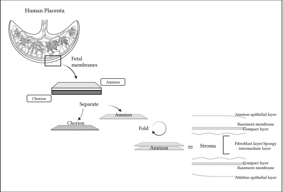
Figure 1: Axolotl DualGraft™ Construction Schematic: An illustration of the fetal membrane layers, showing the amnion and chorion in their native configuration, separated, followed by amnion folding. A detailed cross-section highlights the amnion’s new configuration following folding, with the outer-facing layers being the amnion epithelium followed by the basement membrane, compact layer, and the fibroblast/spongy layer that makes up the stroma (created using BioRender - 2026).
Against this background, the AXOLCAMP study was designed to evaluate the product-specific clinical performance of Axolotl DualGraft™ when used as an adjunct to protocol-defined standard of care in patients with non-healing DFUs. The primary objective is to determine the efficacy of dhAM plus SOC versus SOC alone in achieving complete closure of non-healing DFUs over 12 weeks. Secondary objectives include time to closure over the 12 weeks, percent area reduction (PAR), adverse events relative to SOC alone, and improvement in patient-reported quality of life as measured by the Pain, Enjoyment of Life, and General Activity Scale (PEG), Forgotten Wound Score (FWS), and standard Wound Quality of Life (wQOL) questionnaires. Exploratory objectives include the proportion of ulcers that heal in patients aged 65 and older, and the effect of chronic inhibitory bacterial load (CIBL), assessed by fluorescence imaging at treatment visits 1, 4, 8, and 12. Importantly, this mid-study evaluation specifically evaluates Axolotl DualGraft™ within a randomized, controlled, multicenter framework rather than extrapolating efficacy from the broader placental-tissue literature, providing an opportunity to determine whether this specific dhAM allograft translates into measurable clinical benefit relative to standard care alone.
Methods
Study Design & Registration
AXOLCAMP is a prospective, multicenter, randomized, open-label, controlled clinical trial comparing dehydrated human amnion membrane (dhAM; Axolotl DualGraft™) applied in conjunction with standard of care, with standard of care alone, in patients with nonhealing diabetic foot ulcers. The trial is being conducted under Protocol AXOLOTL1001 and is registered on ClinicalTrials.gov (NCT06550596). A concurrent randomized control group permitted the effect of dhAM to be estimated against an active SOC comparator under standardized conditions. This report presents a mid-study analysis of data provided by the contract research organization (CRO) on 23 September 2026.
Ethics and Informed Consent
The AXOLCAMP study (Protocol AXOLOTL1001; IRB Study No. Pro00080823) was approved by Advarra Institutional Review Board (IRB #00000971) on July 24, 2024, and continues under ongoing reporting and required renewals. All participants provided written informed consent before participation. The study was conducted in accordance with the approved protocol, applicable U.S. federal regulations, and Good Clinical Practice (GCP), with HIPAA requirements maintained and monitored by the CRO, SerenaGroup.
Study Sites
Participants were enrolled at 21 participating wound care centers in the United States, drawn from up to 30 SerenaGroup-affiliated or participating centers eligible for study activation. Sites were clinical practices experienced in DFU management with the infrastructure for prospective wound care research. Before enrolling participants, each site completed IRB authorization, investigator and personnel qualification, protocol and study procedure training, and completion of required study documentation. Each participating site operated under the centrally approved AXOLOTL1001 protocol and associated study documents. Site investigators confirmed participant eligibility, obtained informed consent, oversaw protocol-directed treatment, evaluated the target ulcer, documented outcomes and adverse events, and maintained source and regulatory records. All sites followed the same eligibility criteria, treatment schedule, wound-assessment procedures, offloading requirements, and data collection procedures. Wound management outside of the randomized study intervention was performed according to protocol-defined SOC requirements. Clinical study operations and site coordination were supported by SerenaGroup, Inc., including site and investigator coordination, study conduct support, data collection, and AE/SAE (adverse events/serious adverse events) documentation and escalation. Since AXOLCAMP was designed as a multicenter pragmatic evaluation within participating wound care practices, individual centers could differ in clinical setting, patient referral patterns, and local wound care workflows; however, eligibility, randomized treatment assignment, protocol-defined assessments, and study endpoints were standardized across participating sites.
Participants
Eligible participants were adults (≥18 years) with type 1 or type 2 diabetes mellitus and a qualifying DFU present for 4 to 52 weeks under standard care before the initial screening visit, meeting protocol-defined criteria for wound chronicity, location, severity, size, and vascular sufficiency. At enrollment, the target ulcer measured 1.0 to 20.0 cm² after debridement, to be located on the foot with at least 50% of its area below the malleolus, classified as Wagner grade 1 or 2, and extended at least through the dermis or subcutaneous tissue with no exposed tendon or bone. Adequate limb perfusion was confirmed within 3 months before screening by at least one of the following: ankle-brachial index 0.7 to 1.3, toe-brachial index ≥0.6, transcutaneous oxygen measurement ≥40 mmHg, or a biphasic pulse volume recording. When participants had multiple ulcers, ulcers had to be at least 2 cm apart, with the largest eligible ulcer being designated the target. Participants had to consent to the prescribed offloading method and agree to attend weekly study visits. Following a protocol amendment, 23 September 2024, the allowable target-ulcer area at randomization was expanded from a maximum of 5.0 cm² to 20.0 cm², while maintaining a minimum eligible wound area of 1.0 cm².
The following Table 1 indicates the inclusion and exclusion criteria for the AXOLCAMP study. Deviations from the inclusion and exclusion criteria may occur in special circumstances, to protect the life or physical wellbeing of a subject, with approval from the sponsor, operational manager, and the investigator. Any deviations that may have occurred were accompanied by a protocol deviation form. In addition to these inclusion and exclusion criteria, a subject may exit or discontinue the study voluntarily at any point.
Table 1: Eligibility criteria for AXOLCAMP (Protocol AXOLOTL1001,v3)
|
Inclusion Criterion |
Exclusion Criterion |
|
I1) ≥18 years |
Ulcer & infection: |
|
I2) Type 1 or 2 diabetes mellitus |
Healing during run-in: |
|
I3) Target ulcer area 1.0 to 20.0 cm2 at enrollment, measured after |
Medication & prior therapies: |
|
I4) Target ulcer present for 4 to 52 weeks of SOC before initial |
Comorbidities & Other: |
|
I5) Target ulcer located on the foot with ≥50% of its area below |
|
|
I6) Wagner Grade 1 or 2, extending at least through the dermis |
|
|
I7) Adequate limb perfusion, confirmed within 3 months before |
|
|
I8) If multiple ulcers are present, they must be at least 2 cm apart; |
Subjects may exit or discontinue the study voluntarily at any |
|
I9) Agrees to use the prescribed offloading method for the |
|
|
I10) Agrees to attend the weekly study visits |
|
|
I11) Willing and able to provide informed consent |
|
|
*Amendment from 5.0 to 20.0 cm^2. |
|
Randomization & Blinding
Participants who met all eligibility requirements were randomized in a 1:1 allocation to receive either Axolotl DualGraft™ plus protocol-defined standard of care or standard of care alone. Randomization was performed electronically utilizing Greenlight Guru Clinical data capture system. Since the dhAM application is inherently visible during treatment, masking was not feasible, and the study was conducted under an open-label allocation. Both groups received uniform wound-management procedures in accordance with the protocol, allowing the incremental effect of the dhAM intervention to be evaluated against a rigorously applied standard-of-care comparator. This randomized controlled design was intended to reduce the influence of baseline wound heterogeneity, site-specific practice variation, and other clinical factors known to influence diabetic foot ulcer healing trajectories.
Subjects were followed longitudinally throughout the treatment period with serial wound assessments and standardized collection of clinical outcomes. The primary efficacy endpoint was the proportion of target ulcers achieving complete wound closure during the defined treatment period. Secondary outcomes included time to closure, percent area reduction (PAR), adverse events (AEs), pain-related measures, and wound-related quality-of-life measures. Wound progression and safety were assessed at each scheduled study visit according to protocol-defined procedures. The multicenter design was intended to broaden the clinical relevance of the findings while preserving consistency in subject eligibility criteria, wound assessment techniques, treatment procedures, and outcome documentation across participating sites. All study procedures were conducted in accordance with the protocol, applicable GCP principles, institutional requirements, and relevant U.S. regulatory standards.
Interventions
Both groups received protocol-defined SOC, consisting of debridement, reduction of bacterial burden, and maintenance of moist balance and offloading. Participants randomized to the dhAM group received up to eight applications of Axolotl DualGraft™ in addition to SOC; SOC alone was provided at protocol-defined visits when dhAM was not applied; see Chart 1 for a breakdown of participants’ visits.
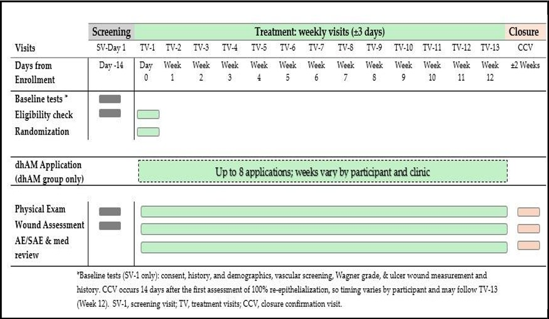
Chart 1: AXOLCAMP Trial Visit Schedule (Protocol AXOLOTL1001, v3, 2024): Timeline of the study visits for participants, beginning with the screening, progressing through the 12 weeks of treatment and ending with closure confirmation. The chart outlines the baseline assessments, dhAM application windows, and some of the recurring evaluations that take place at each visit, including the physical exam, wound assessment, and safety monitoring.
The dhAM product (Axolotl DualGraft™, Axolotl Biologix, Scottsdale, AZ, USA) was derived from full-term human placental tissue donated following live birth by consenting maternal donors. Donor eligibility, tissue recovery, processing, and associated quality activities were conducted through an Association for Advancing Tissue and Biologics (AATB) accredited and U.S. Food and Drug Administration (FDA) registered tissue bank operating in accordance with applicable AATB standards and 21 CFR Part 1271 governing human cells, tissues, and cellular and tissue-based products (HCT/Ps) [13, 14]. Placental tissue was obtained following documented maternal consent and processed under established tissue bank procedures for donor screening, tissue recovery, handling, traceability, and quality oversight. Following recovery, the amnion was mechanically dissected from the chorion and processed as a minimally manipulated human tissue allograft. The isolated amniotic membrane was configured as a dual-layer construct with the stromal surfaces oriented inward, and amniotic epithelial surfaces facing outward on both sides. Dehydration was performed under controlled processing conditions followed by terminal sterilization using a validated gamma-irradiation process, and supplied as a sterile, dehydrated graft stored at ambient temperature. The product sizing, application, storage, and accountability were in accordance with applicable HCT/P requirements and the study protocol.
Study Procedures and Outcomes
Randomized participants were scheduled for weekly study visits for up to 12 weeks (TV1 = Week 0, TV13 = Week 12) and underwent serial wound assessments and standardized collection of clinical outcomes. Wounds judged closed were to be re-examined 2 weeks later to confirm and re-evaluate closure. The primary efficacy outcome was the proportion of target ulcers achieving complete wound closure within 12 weeks, defined as full re-epithelialization without drainage, confirmed at the 2-week visit. Secondary outcomes were time to closure, percent area reduction, adverse events, pain score, and the wound-related quality of life. Safety and tolerability were monitored throughout participation.
Study Oversight & Operational Conduct
AXOLCAMP was sponsored by Axolotl Biologix, with SerenaGroup Inc. serving as the contract research organization (CRO) responsible for site selection and coordination, clinical operations, data collection, documentation, and escalation of adverse events (AEs) and serious adverse events (SAEs) in accordance with the study protocol and applicable regulatory requirements. The present mid-study analysis was initiated, evaluated, and led by the study sponsor using data generated within the ongoing clinical trial under the supervision of the designated Medical Director and participating site investigators. Under the clinical study agreement, SerenaGroup was required to promptly notify the Sponsor of serious adverse events and other information that could affect participant safety, medical care, study conduct, or data integrity. Safety information generated at participating sites was therefore managed through the CRO-led clinical operations structure, with Sponsor oversight maintained throughout the study. Study activities were performed in accordance with the protocol, applicable U.S. federal and state requirements, International Council for Harmonization Good Clinical Practice (ICH-GCP) principles, HIPAA requirements, and applicable institutional policies practiced by SerenaGroup, Inc. This mid-study analysis was initiated, led, and interpreted by the sponsor using data from the ongoing CRO-executed controlled clinical trial. All subject-facing clinical activities and site-level study procedures were conducted through SerenaGroup and its participating clinical sites under the approved protocol while the sponsor remained responsible for study sponsorship and the interpretation of the mid-study dataset.
Statistical Analysis
The data presented are from a mid-study analysis and reflect ongoing data collection and patient participation, using data provided on 23 September 2026. Statistical analysis was completed in RStudio (Version 2024.12.0) and GraphPad Prism (Version 10.3.1). Artificial intelligence (AI) was used for data cleaning, compilation, and preparation of analysis-ready datasets (OpenAI). AI-assisted outputs were individually reviewed by the authors for accuracy. The outcomes reported in this mid-study analysis include complete wound closure, percent area reduction (PAR), adverse events (AE), and pain assessed utilizing the Pain, Enjoyment, and General Activity (PEG) scale. Other protocol-defined outcomes, including time to closure, quality of life assessment, forgotten wound scores, and bacterial assessments, are outside the scope of this mid-study report.
Importantly, complete wound closure in this analysis was modified from the protocol’s prespecified criteria. Subjects marked as “complete” who also reached a final wound area size of 0 cm2 were deemed full closure, regardless of 2-week follow-up confirmation. The decision to modify the protocol’s specified requirements was made preemptively because of the nature of this data set. Many subjects were ongoing, recently completed, or awaiting 2-week follow-up, which could have excluded subjects with meaningful observations or restricted the available sample size if the definition was not modified to reflect the ongoing nature of the study.
Subjects were summarized according to their randomized treatment group, standard of care (SOC), or SOC + Treatment. Continuous variables were described using means ± standard deviations (SD) or medians and interquartile ranges (IQR) as appropriate. Categorical variables were described using counts and percentages. Continuous variables were compared between treatment groups using two-sided Welch’s t-tests or Wilcoxon tests, and categorical variables were compared using Fisher’s exact tests. An exploratory linear mixed-effects model was conducted on the longitudinal PAR data with treatment group as a categorical variable and study week modeled as a continuous variable, and their interaction included as a fixed effect. Subject-specific random intercept and slopes were included. Models were fitted using maximum likelihood and no multiplicity adjustment was used. The interaction coefficient represented the between-group difference in PAR change as percentage points per week. Between-group differences in PEG score change were evaluated among patients with both a baseline and TV13 measurement.
Missing values were not imputed, and analysis of continuous variables used only available observations. Participants who did not reach the relevant assessment at the cutoff contributed only their earlier measurements, and unavailable assessments were not assigned a value of zero. All reported p-values are two-sided. The visit-specific and longitudinal comparisons were exploratory and the p-values should be interpreted as such.
Results
The CRO supplied data to the sponsor on 23 September 2026, as of that timepoint a total of 171 subject records were entered into the study database across 21 unique sites. Of those, 86 subjects have been assigned a randomized protocol group, while the remaining 85 subjects were excluded, discontinued, or are in the pre-randomization stage. A full breakdown of subject enrollment and status through the study is provided in the CONSORT flow diagram (Figure 2).
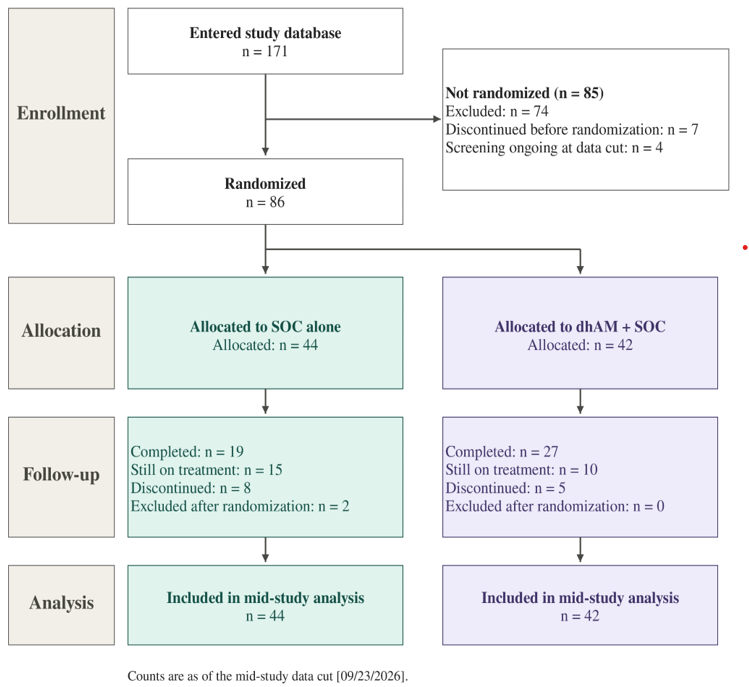
Figure 2: CONSORT flow diagram for AXOLCAMP mid-study analysis, summarizing participant progression from database entry, through randomization, treatment allocation, follow-up, and inclusion in the mid-study analysis. The counts reflect the current enrollment status, exclusions, discontinuations, and ongoing treatment at the mid-study data cut date of September 23rd 2026.
Of the 86 subjects randomized into treatment groups, there were no significant differences in demographics (age, sex, race, etc.) and no significant differences in baseline wound characteristics (age, size, Wagner grade, etc.). The majority of patients were male (73.3%) and under the age of 65 (77.9%). Subject demographics and baseline wound characteristics are summarized in Table 2.
Table 2: Subject demographics and baseline wound characteristics for randomized participants (n=86). The data is presented as mean ± standard deviation, as a percentage, or as median (interquartile range [IQR]), as appropriate. Between-group comparisons were performed using independent-samples t-test for normally distributed continuous variables, Wilcoxon rank-sum test for non-normally distributed continuous variables, and Fisher’s exact test for categorical variables. MNA = mini nutritional assessment.
*HbA1C % could be taken offsite or onsite; when available, the onsite metric was used for analysis; otherwise, offsite reported metrics were used. One subject reported HbA1C % value was identified as implausible and was omitted (107.1).
|
Characteristic |
Level |
Overall (n=86) |
SOC (n=44) |
Treatment + SOC (n=42) |
p-value |
|
Age, Years |
|
57.4 ± 12.2 |
58.6 ± 11.2 |
56.2 ± 13.2 |
0.351 |
|
Age Group |
<65 |
67 (77.9%) |
34 (77.3%) |
33 (78.6%) |
1.000 |
|
≥65 |
19 (22.1%) |
10 (22.7%) |
9 (21.4%) |
|
|
|
Sex |
Male |
63 (73.3%) |
29 (65.9%) |
34 (81.0%) |
0.146 |
|
Female |
23 (26.7%) |
15 (34.1%) |
8 (19.0%) |
|
|
|
BMI, kg/m2 |
|
32.9 ± 7.8 |
34.0 ± 8.8 |
31.8 ± 6.3 |
0.186 |
|
HbA1c, %* |
|
8.1 ± 2.2 |
8.1 ± 2.3 |
8.0 ± 2.0 |
0.822 |
|
MNA Score |
|
25.0 ± 2.8 |
25.1 ± 2.8 |
25 ± 2.8 |
0.765 |
|
Ethnicity |
Hispanic/Latino |
20 (23.3%) |
10 (22.7%) |
10 (23.8%) |
1.000 |
|
Non-Hispanic/Latino |
66 (76.7%) |
34 (77.3%) |
32 (76.2%) |
|
|
|
Race |
Asian |
1 (1.2%) |
1 (2.3%) |
0 (0.0%) |
0.658 |
|
Black |
14 (16.3%) |
6 (13.6%) |
8 (19.0%) |
|
|
|
Hawaiian/Pacific Islander |
1 (1.2%) |
0 (0.0%) |
1 (2.4%) |
|
|
|
Other |
5 (5.8%) |
2 (4.5%) |
3 (7.1%) |
|
|
|
White |
65 (75.6%) |
35 (79.5%) |
30 (71.4%) |
|
|
|
Tobacco User |
Yes |
12 (14.0%) |
5 (11.4%) |
7 (16.7%) |
0.545 |
|
No |
74 (86.0%) |
39 (88.6%) |
35 (83.3%) |
|
|
|
Wound Characteristics |
|||||
|
DFU Age at |
|
19.5 (10.0-36.0) |
16.0 (8.0-33.0) |
20.5 (11.5-37.5) |
0.481 |
|
Screening Wound |
|
3.0 (1.6-5.4) |
2.6 (1.6-4.3) |
3.2 (1.7-6.9) |
0.144 |
|
Baseline Wound |
|
0.3 (0.2-0.4) |
0.3 (0.2-0.4) |
0.3 (0.2-0.4) |
0.567 |
|
Wagner Grade |
Grade 1 |
63 (73.3%) |
30 (68.2%) |
33 (78.6%) |
0.334 |
|
Grade 2 |
23 (26.7%) |
14 (31.8%) |
9 (21.4%) |
|
|
|
Baseline Bacterial |
Positive |
41 (49.4%) |
19 (44.2%) |
22 (55.0%) |
0.383 |
|
|
Negative |
42 (50.6%) |
24 (55.8%) |
18 (45.0%) |
|
The primary endpoint for this study is defined as the number and percentage of subject target ulcers that reach full closure within 12 weeks. Importantly, because this is a mid-study analysis of an ongoing trial, full closure of a target ulcer was defined as any subject who is marked as “complete” who also reached a wound size of 0 cm2 within this timeframe. The SOC + Treatment group had a full wound closure percentage of 18.5% compared to 15.8% in the SOC alone group. The results are summarized in Table 3 and are intended as exploratory, as the study is still ongoing.
Table 3: Wound-closure outcome, number and percentage of participants with apparent full wound closure within 12 weeks among participants with evaluable follow-up. For this mid-study analysis, apparent full wound closure was defined as any subject marked completed at the data cutoff point who reached a wound area of 0 cm2.
|
Group |
Closed |
Completed |
Closure % |
|
Standard of Care |
3 |
19 |
15.8% |
|
Treatment + Standard of Care |
5 |
27 |
18.5% |
One of the secondary endpoints for this study is percent area reduction (PAR) for target ulcers (Figure 3). PAR is calculated as a percentage decrease in wound area from baseline (TV1, week 0): [(TV1 area - follow-up area) / TV1 area] x 100. A general trend of increased PAR in the SOC + Treatment compared to SOC alone can be observed in Figure 3. At TV2, SOC and SOC + Treatment mean PAR were 9.39 and 7.70, respectively. At TV8, mean PAR was 21.77 and 37.65, respectively. Finally, by TV13, mean PAR was 19.16 and 41.07, respectively. Despite the observable trend, between-group comparisons at any given week did not reach statistical significance. Between-group comparisons are summarized in Table 4. Importantly, PAR was calculated using all recorded observations available for each group at the study week. As this is an ongoing study n values for each study week and treatment group varied as missing data was not imputed.
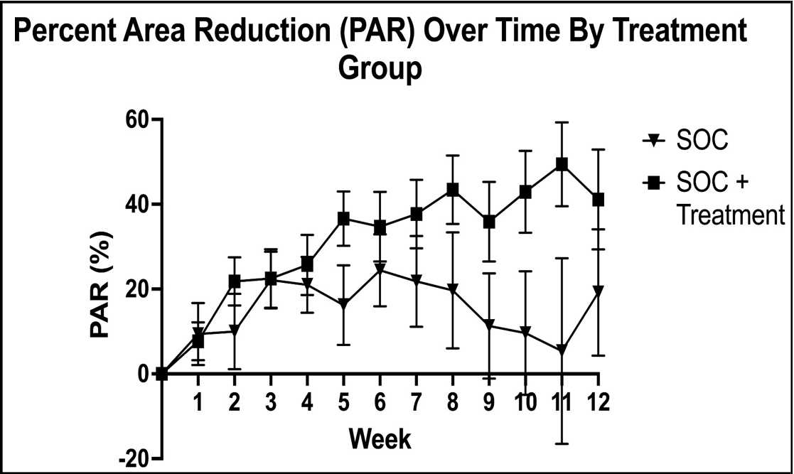
Figure 3: Secondary endpoint, percent area reduction, over time by treatment group. Mean PAR values are shown for SOC and SOC + Treatment groups from baseline week 0 (TV1) through week 12 (TV13). PAR was calculated relative to TV1 wound area, with positive values indicating wound size reduction and negative values indicating wound area enlargement. Data is presented as mean ± SEM using all available observations at each visit. Sample size varies between visits and groups as missing values were not imputed.
Table 4: Between-group comparisons at individual visit marks. Comparison performed using a two-sided Welch’s t-test; no visit reached statistical significance. Values are mean PAR (%) for SOC and SOC + Treatment groups. No adjustment for multiple comparisons was applied, p-values are exploratory.
|
Visit |
SOC mean PAR (%) |
SOC + Treatment PAR (%) |
p-value |
|
TV2 |
9.39 (n = 41) |
7.70 (n = 39) |
0.844 |
|
TV3 |
10.00 (n = 37) |
21.76 (n = 37) |
0.272 |
|
TV4 |
22.05 (n = 37) |
22.49 (n = 33) |
0.963 |
|
TV5 |
20.96 (n = 37) |
25.69 (n = 34) |
0.627 |
|
TV6 |
16.17 (n = 35) |
36.58 (n = 33) |
0.079 |
|
TV7 |
24.36 (n = 31) |
34.75 (n = 30) |
0.383 |
|
TV8 |
21.77 (n = 28) |
37.65 (n = 30) |
0.241 |
|
TV9 |
19.68 (n = 25) |
43.31 (n = 29) |
0.146 |
|
TV10 |
11.30 (n = 24) |
35.90 (n = 29) |
0.120 |
|
TV11 |
9.64 (n = 24) |
42.92 (n = 27) |
0.065 |
|
TV12 |
5.41 (n = 20) |
49.44 (n = 23) |
0.078 |
|
TV13 |
19.16 (n = 21) |
41.07 (n = 26) |
0.255 |
As an exploratory longitudinal analysis, a linear mixed-effect model was used to evaluate longitudinal PAR, utilizing treatment group, study week, and treatment-by-week interaction as fixed effects. Subject-specific random intercepts and slopes were used to account for repeated measurements. The analysis included 730 post-baseline observations from 82 participants across week 1 through 12. The models were fitted utilizing maximum-likelihood fitting and a likelihood-ratio comparison of models with and without the treatment-by-week interaction. In the SOC group, PAR increased by an estimated 0.74 percentage points per week, while the SOC + Treatment group increased an estimated 4.07 percentage points per week. This model demonstrated a 3.33 percentage point increase in PAR per week in the SOC + Treatment group compared to SOC alone (β = 3.33, SE = 1.67, 95% CI = 0.06-6.60). Addition of the treatment-by-week interaction significantly improved the model fit (p = 0.049). Additional secondary endpoints evaluated in this study include number of adverse events and pain and quality of life scorings. Both treatment groups had 14 subjects with at least 1 adverse event reported at the data collection cutoff point. Mean PEG scores at TV13 differed significantly between groups (SOC = 2.86 ± 2.97 (n=23), SOC + Treatment = 1.31 ± 2.11 (n=26), p = 0.044), while the mean change from TV1 to TV13 was not significant (p = 0.774). A lower PEG score represents less pain-related interference. These results are presented collectively in Table 5. A decrease in PEG observations from baseline to TV13 was due to multiple factors including ongoing subjects yet to reach week 12, discontinuations, withdrawals, and incomplete documentation.
Table 5: Secondary and exploratory mid-study outcomes by treatment group. Adverse events are reported as a percentage of randomized subjects per group with at least one reported AE/SAE. PEG scores are reported as mean ± SD; lower scores indicate less pain-related interference. The protocol-defined PEG endpoint was change from baseline (TV1) to TV13; analysis includes only subjects who have both a baseline and TV13 score reported. Baseline and TV13 scores are independently reported for subjects who have either score at that timepoint. Between-group comparisons were performed using a two-sided Welch’s t-test and categorical outcomes were compared using Fisher’s exact test. Sample sizes vary by outcome and visit number because of the ongoing nature of the study.
|
Outcomes |
Timepoint/Metric |
SOC |
SOC + Treatment |
p-value |
|
Adverse events |
Participants with ≥1 AE/SAE, n/N (%) |
14/44 (31.8%) |
14/42 (33.3%) |
1.000 |
|
PEG score |
TV1 baseline, mean ± SD |
2.73 ± 2.52 (n=44) |
1.90 ± 2.41 (n=42) |
0.126 |
|
TV13, mean ± SD |
2.86 ± 2.97 (n=23) |
1.31 ± 2.11 (n=26) |
0.044 |
|
|
Change TV1 to TV13, mean ± SD |
−0.23 ± 2.22 (n=23) |
−0.06 ± 1.78 (n=26) |
0.774 |
Discussion
Diabetic foot ulcers remain among the most clinically consequential complications of diabetes, with prolonged healing, recurrent ulceration, infection, hospitalization, lower-extremity amputation, and mortality contributing substantially to the individual and health-system burden of disease [3]. Despite advances in multidisciplinary wound care, a substantial proportion of DFUs fail to achieve timely closure with standard treatment alone. Contemporary guidelines emphasize comprehensive management that includes regular wound assessment, appropriate debridement, pressure offloading, vascular evaluation, infection management, moisture balance, and optimization of systemic factors; however, adjunctive wound-healing interventions may be considered when appropriately delivered standard of care is insufficient to achieve adequate healing progression [15]. This persistent therapeutic gap provides the rationale for evaluating advanced tissue-based interventions which complement, rather than replace, established wound-care practices. Cellular and acellular matrix products (CAMPs), including amniotic membrane allografts, have gained increasing attention as adjunctive treatments for intractable DFUs. Randomized clinical trials and systematic reviews evaluating several amniotic and amnion/chorion products have demonstrated improved healing outcomes compared with SOC alone in selected patient populations [9, 10, 16]. However, these products are not biologically or structurally interchangeable. Tissue source, membrane composition, processing, preservation, sterilization, configuration, and clinical application vary across products and may influence clinical performance [4].
AXOLCAMP was designed to evaluate a dehydrated, dual-layer, chorion-free human amnion membrane (Axolotl DualGraft™) as an adjunct to protocol-defined SOC in chronic, nonhealing DFUs. This mid-study analysis reflects early findings of the intervention within an ongoing prospective, randomized, multicenter trial conducted across 21 wound-care centers. At the current data cutoff, 86 participants had been randomized, with baseline demographic and wound characteristics generally comparable between groups.
The primary objective of AXOLCAMP is complete target-ulcer closure during the 12-week treatment period. At this data cutoff, apparent complete closure was observed in 18.5% of evaluable participants receiving dhAM plus SOC compared with 15.8% receiving SOC alone, indicating a modest early numerical advantage for adjunctive dhAM treatment. These proportions remain preliminary, as a substantial proportion of randomized participants had not yet completed the full 12-week observation period, and therefore have not had the protocol-defined opportunity to achieve closure. Within this context, the directional advantage observed in the dhAM plus SOC group is encouraging, particularly when considered alongside the progressively greater PAR noted at later study visits.
Whether this early difference in complete closure expands, persists, or attenuates as additional participants complete follow-up will be determined in the prespecified final analysis after completion of enrollment and the full observation period.
While complete wound closure remains the clinically definitive endpoint, longitudinal changes in wound area may provide early insight into treatment response before sufficient closure events accumulate. In this mid-study analysis, one of the more notable observations was the progressively greater separation in PAR between treatment groups at later visits. Early post-randomization changes were generally similar; however, by week 12, mean PAR was 41.07% in the dhAM plus SOC group compared with 19.16% among those receiving SOC alone. Although individual visit-level comparisons did not reach statistical significance, the consistent directional pattern observed across later visits suggests that the overall trajectory of wound reduction may provide additional information beyond isolated cross-sectional comparisons at individual timepoints.
This interpretation was further supported by the exploratory longitudinal mixed-effects analysis, which accounted for repeated measurements within participants and evaluated differences in the rate of wound-area reduction over time. In this model, PAR increased by an estimated 0.74 percentage points per week in the SOC group compared with 4.07 percentage points per week with adjunctive dhAM. The treatment-by-week interaction corresponded to an additional 3.33 percentage-point increase in PAR per week in the treatment group (β = 3.33, SE = 1.67), and inclusion of the interaction significantly improved model fit (p = 0.049). Although exploratory and near the conventional threshold for statistical significance, this finding represents a longitudinal signal that warrants confirmation in the completed dataset. This model does not establish a significant difference in PAR at any specific week, rather it suggests a change in healing trajectories. Taken together with the later visit-level PAR differences, these data raise the possibility that adjunctive dhAM may influence the trajectory of wound reduction progressively over the treatment period rather than producing an immediate separation between groups. Complete epithelialization represents the culmination of coordinated processes that include wound-bed preparation, extracellular matrix remodeling, granulation tissue formation, cellular migration, and re-epithelialization. Changes in wound dimensions may therefore precede measurable differences in complete closure. Early wound-area reduction has been established as a useful prognostic indicator of subsequent DFU healing, with greater reduction during the first several weeks of treatment associated with a higher probability of later complete wound closure [17]. PAR should, however, be considered a measure of healing progression rather than a substitute for the clinically definitive endpoint of complete wound closure.
The mid-study safety findings are an important consideration for an adjunctive treatment intended for repeated use in chronic wound management. The proportion of randomized participants experiencing at least one adverse event or serious adverse event was similar between groups, occurring in 31.8% of participants receiving SOC alone and 33.3% receiving dhAM plus SOC. This mid-study safety pattern is generally consistent with prior randomized studies of amniotic-derived products, in which overall adverse-event incidence was comparable between treatment and SOC groups [18]. At the current cutoff, the overall proportion of participants with reported adverse events was similar between groups; however, these descriptive findings should not be interpreted as establishing safety equivalence. However, this observation remains preliminary, as event adjudication and classification are ongoing. The overall incidence of participants experiencing an event does not distinguish event severity, seriousness, relationship to study treatment, or clinically relevant wound complications, all of which will be clarified in the completed dataset.
The patient-reported findings provide an additional and potentially encouraging perspective on treatment response beyond objective wound measurements. At TV13, participants receiving dhAM plus SOC reported significantly lower mean PEG pain-interference scores than participants receiving SOC alone (1.31 ± 2.11 versus 2.86 ± 2.97; p = 0.044), indicating less pain-related interference among participants contributing data at this later study timepoint. This observation is clinically relevant because pain associated with diabetic foot ulceration has been linked to poorer health-related quality of life and limitations in physical functioning, while mobility restrictions and patient-related factors can also influence adherence to prescribed offloading regimens [19, 20]. However, the between-group difference in change from baseline to TV13 was not statistically significant [SOC = −0.23 ± 2.22 (n = 23) vs. SOC + Treatment = −0.06 ± 1.78 (n = 26); p = 0.774]. The TV13 and baseline to TV13 comparison analysis may be affected by which subjects remained within the analysis group, as only subjects who reached TV 13, or had both a baseline and TV13 observation were included. Because enrollment and follow-up remain ongoing, variable study completion and missing patient-reported assessments may also contribute to uncertainty in the current estimates. Taken together, the significantly lower PEG score observed at TV13 represents a favorable patient-reported signal, while paired observations remain not significant, this warrants further evaluation as the trial matures. Although the present findings do not establish that dhAM independently reduces pain, they raise the possibility that improvements in wound progression, local wound condition, or treatment experience may translate into reduced pain-related interference over time.
The current observations are generally consistent with the broader clinical rationale for evaluating amniotic-derived allografts as adjunctive therapies in chronic wound management. Prior prospective randomized studies have demonstrated improved DFU healing and complete closure with selected amniotic and amnion/chorion preparations when used in addition to standard of care (SOC) [10, 18, 21]. Systematic review and meta-analytic evidence has similarly demonstrated a greater likelihood of complete healing with human amniotic membrane products compared with SOC alone across randomized studies [16]. These findings support the clinical relevance of placental-derived tissue products as a therapeutic category. In addition, differences in study eligibility criteria, baseline wound characteristics, offloading practices, treatment frequency, endpoint definitions, and approaches to missing data complicate direct comparison across clinical trials. The importance of AXOLCAMP therefore lies in its prospective evaluation of this specific dual-layer, chorion-free dhAM construct against a concurrent randomized SOC comparator rather than reliance on class-level evidence.
Recent multicenter RCTs have further demonstrated the feasibility and importance of evaluating placental-derived products across diverse wound-care settings. Tettelbach et al. evaluated dehydrated human amnion/chorion membrane across 14 wound-care centers, while Cazzell et al. recently reported a randomized multicenter study of dehydrated amnion/chorion membrane in a comparatively complex DFU population [18, 21]. In the latter study, complete wound closure at 12 weeks occurred more frequently with the placental-derived intervention plus SOC than with SOC alone, further supporting the importance of product-specific randomized evaluation in clinically heterogeneous populations [21]. AXOLCAMP extends this approach across 21 participating wound-care centers. Although multicenter execution introduces expected variability in referral patterns, patient characteristics, and routine clinical workflows, core eligibility criteria, randomized treatment assignment, outcome definitions, scheduled assessments, and protocol-directed study procedures were standardized across participating sites. This design therefore provides an important balance between methodological control and clinical applicability and may increase the relevance of the eventual findings to outpatient wound-care practice.
An additional consideration when interpreting AXOLCAMP is that the study intentionally selected a population with wounds demonstrating limited improvement during preceding SOC. Participants whose target ulcers decreased in area by 20% or more during the defined screening period were excluded from randomization. Thus, the randomized population was enriched for ulcers demonstrating inadequate short-term progression despite standardized wound management. This is clinically relevant because early wound-area reduction is a recognized prognostic indicator of subsequent DFU healing; greater reductions during the first several weeks of care are associated with a higher probability of later complete closure [17]. Similarly, AXOLCAMP evaluates adjunctive dhAM in a particularly relevant and therapeutically challenging population rather than in an unselected cohort of newly presenting DFUs. This characteristic should be considered when comparing absolute closure rates between AXOLCAMP and studies enrolling wounds with greater spontaneous healing potential.
The AXOLCAMP protocol amendment expanding the maximum permitted target-ulcer area from 5.0 cm² to 20.0 cm² further broadened the range of wound complexity represented in the study. Baseline wound area is an established prognostic factor in DFU healing, with larger wounds generally demonstrating a lower probability of closure or requiring longer periods to heal than smaller wounds [22]. Wound duration and baseline area have also been shown to remain important predictors of healing across contemporary DFU cohorts [22]. Inclusion of larger target ulcers may therefore make the AXOLCAMP population more representative of the spectrum of wounds encountered in clinical practice, while simultaneously increasing heterogeneity in healing potential. The completed analysis should therefore evaluate whether baseline wound size modifies treatment response and whether the observed treatment effects remain consistent after adjustment for wound area and when participants enrolled before and after the protocol amendment are considered separately.
Taken together, the mid-study findings provide an encouraging signal regarding the potential clinical performance of Axolotl DualGraft™ when used as an adjunct to standard of care. In particular, the progressively greater PAR observed during later study visits, together with the favorable treatment-by-time interaction in the exploratory longitudinal analysis and the absence of an apparent increase in overall adverse-event burden, support continued evaluation of the product within the ongoing randomized trial. Although these findings are not yet sufficient to establish definitive clinical efficacy, they demonstrate promising trends consistent with a potential treatment-associated improvement in wound-healing progression. The final determination of clinical benefit will depend on completion of enrollment, maturation of the primary wound-closure endpoint, and the prespecified analysis of the full study population. If the favorable longitudinal healing pattern observed in the present mid-study analysis is maintained and translates into greater complete and durable wound closure, the completed AXOLCAMP trial may provide important product-specific evidence supporting the use of this dual-layer dhAM allograft as an adjunct to comprehensive standard care for nonhealing diabetic foot ulcers.
Limitations
Several limitations should be considered when interpreting this mid-study analysis. Most importantly, AXOLCAMP remains ongoing, and the present dataset includes participants who completed treatment, discontinued participation, or remained under active follow-up at the data cutoff. Accordingly, outcome maturity and available sample size vary across endpoints and visits. The descriptive PAR analyses used available observations without imputation and missing data may influence visit-specific estimates. Because follow-up was incomplete at the data cutoff, differential attrition or patterns of missing observations between treatment groups could also introduce bias into the interim estimates. In addition, the limited number of complete closure events precludes definitive conclusions regarding the primary endpoint or time to closure at this stage of the trial.
Because of the physical nature of the intervention, AXOLCAMP is open-label and treatment allocation cannot be practically concealed from investigators or participants. Randomization, standardized eligibility criteria, protocol defined wound assessments, and standardized treatment procedures reduce, but do not eliminate, the potential influence of this limitation. The multicenter design may also introduce variability in patient populations, wound-care practices, offloading adherence, debridement, and other site-level factors despite protocol standardization. In addition, several analyses presented in this mid-study evaluation, including the longitudinal mixed-effects assessment of PAR, are exploratory and require confirmation in the completed dataset using the prespecified statistical analysis plan. Final analyses should also fully characterize attrition, missing outcomes, and protocol deviations. Finally, the study is sponsored by Axolotl Biologix, with Sponsor involvement in the present analysis and manuscript preparation. Clinical trial operations, site coordination, data collection, and safety reporting were conducted through SerenaGroup and participating investigators under the approved protocol.
Conclusion
This mid-study analysis of AXOLCAMP provides preliminary evidence regarding the use of a dehydrated dual-layer human amnion membrane as an adjunct to standard of care for chronic, nonhealing diabetic foot ulcers. At the current data cutoff, the randomized treatment groups remained generally well balanced with respect to baseline demographic and wound characteristics, supporting the integrity of the ongoing comparison. Importantly, adjunctive dhAM treatment was not associated with an apparent increase in the overall proportion of participants experiencing reported adverse events.
Although the modified primary endpoint of apparent complete wound closure remains immature, the evolving wound healing trajectory observed in the treatment group is worth noting. Participants receiving dhAM plus standard of care demonstrated progressively greater percent area reduction during later study visits, and the exploratory longitudinal analysis identified a significant treatment-by-time interaction. Taken together, these findings provide an encouraging trend consistent with a potential treatment associated with improvement in wound healing progression. These mid-study observations should be interpreted as hypothesis generating rather than confirmatory. The current dataset does not yet permit definitive conclusions regarding complete or durable wound closure, time to closure, or overall clinical efficacy; however, the direction and longitudinal pattern of the wound area response support continued evaluation of Axolotl DualGraft™ within the ongoing randomized multicenter trial. Following completion of full study enrollment and protocol defined participant follow-up, a comprehensive final analysis will be performed using the prespecified analysis population and statistical methods. This analysis will determine whether the favorable wound healing trajectory observed in the present mid-study evaluation translates into a clinically and statistically meaningful improvement in complete and durable wound closure and will provide a more definitive assessment of safety, percent area reduction, time to closure, pain, wound-related quality of life, and other prespecified secondary and exploratory outcomes. Overall, the current findings provide a positive trend and scientific foundation for the completed AXOLCAMP analysis and support the continued investigation of this specific dhAM allograft, Axolotl DualGraft™, as an adjunct to the comprehensive standard of care for diabetic foot ulcers.
Acknowledgments
The sponsor gratefully acknowledges SerenaGroup, Inc. for its support of the AXOLCAMP study, along with the patients who enrolled and the families who supported their participation. This study would not have been possible without their time, commitment, and continued support.
Author Contributions
Conceptualization, DC, AI, JS and AJT; methodology DC, AI and AJT; validation, DC; formal analysis, DC, AI and AJT; resources, JS and AJT; data curation, DC and AI; writing—original draft preparation DC, AI and AJT; writing—review and editing, DC, AI, JS and AJT; visualization, DC, AI and AJT; supervision, AJT; project administration, AJT. All authors have read and agreed to the published version of the manuscript.
Funding
This research received no external grant funding. Axolotl Biologix sponsored the AXOLCAMP trial and led the present mid-study analysis and manuscript development. Clinical trial operations, site selection, coordination, and study execution were performed by SerenaGroup, Inc. and participating investigators in accordance with the study protocol. The Sponsor was involved in study oversight, interpretation of the mid-study findings, and preparation of the manuscript.
Institutional Review Board Statement
The AXOLCAMP study (Protocol AXOLOTL1001; IRB Study No. Pro00080823) received initial approval from the Advarra Institutional Review Board on July 24, 2024 with ongoing yearly reports and renewals. Advarra IRB is registered with the U.S. Office for Human Research Protections and the U.S. Food and Drug Administration as IRB #00000971.
Informed Consent Statement
Written informed consent was obtained from all participants prior to enrollment and initiation of study-related procedures. The informed consent process was conducted using the IRB approved consent document for Protocol AXOLOTL1001, which was approved by Advarra IRB. Participation was voluntary, and subjects were informed of the study purpose, procedures, potential risks and benefits, alternative treatment options, privacy protections, and their right to withdraw from the study at any time without penalty.
Data Availability Statement
The datasets present in this article are not readily available because of corporate policy and ongoing studies with dhAM. Requests for access to the data set should be directed to Dr. Aaron J. Tabor
Conflicts of Interest
D.C., A.I., J.S., and A.J.T. are employees of or contractors to Axolotl Biologix. J.S. is the founder and an owner of Medtech Incubator, and A.J.T. is an owner of Medtech Incubator.
Abbreviations
The following abbreviations are used in this manuscript:
AATB Association for Advancing Tissue and Biologics
ABI Ankle-Brachial Index
AE Adverse Event
AI Artificial Intelligence
BMI Body Mass Index
CAMP Cellular, Acellular, or Matrix-like Product
CFR Code of Federal Regulations
CI Confidence Interval
CIBL Chronic Inhibitory Bacterial Load
CONSORT Consolidated Standards of Reporting Trials
CRO Contract Research Organization
DFU Diabetic Foot Ulcer
dhAM Dehydrated Human Amnion Membrane
FDA Food and Drug Administration
FWS Forgotten Wound Score
GCP Good Clinical Practice
GTP Current Good Tissue Practices
HbA1c Glycated Hemoglobin
HCT/Ps Human Cells, Tissues, and Cellular and Tissue-based Products
HIPAA Health Insurance Portability and Accountability Act
ICH-GCP International Council for Harmonization Good Clinical Practice
IQR Interquartile Range
IRB Institutional Review Board
ISO International Organization for Standardization
MNA Mini Nutritional Assessment
PAR Percent Area Reduction
PEG Pain, Enjoyment of Life, and General Activity
PVR Pulse Volume Recording
RCT Randomized Controlled Trial
S1 Screening Visit 1
SAE Serious Adverse Event
SD Standard Deviation
SE Standard Error
SEM Standard Error of the Mean
SOC Standard of Care
TBI Toe-Brachial Index
TCOM Transcutaneous Oxygen Measurement
TV Treatment Visit
wQOL Wound Quality of Life
References
- Senneville É, Albalawi Z, van Asten SA, Abbas ZG, Allison G, et al. (2024) IWGDF/IDSA guidelines on the diagnosis and treatment of diabetes-related foot infections (IWGDF/IDSA 2023). Diabetes Metab Res Rev. 40(3): e3687.
- Ndosi M, Wright-Hughes A, Brown S, Backhouse M, Lipsky BA, et al. (2018) Prognosis of the infected diabetic foot ulcer: A 12-month prospective observational study. Diabet Med. 35(1): 78–88.
- McDermott K, Fang M, Boulton AJM, Selvin E, Hicks CW (2023) Etiology, epidemiology, and disparities in the burden of diabetic foot ulcers. Diabetes Care. 46(1): 209–221.
- Ingraldi AL, Audet RG, Tabor AJ (2023) The preparation and clinical efficacy of amnion-derived membranes: A review. J Funct Biomater. 14(10): 531.
- Fitriani N, Wilar G, Narsa AC, Mohammed AFA, Wathoni N (2023) Application of amniotic membrane in skin regeneration. Pharmaceutics. 15(3): 748.
- Guo X, Zhang W, Lu J, Zhu Y, Sun H, et al. (2024) Amniotic miracle: Investigating the unique development and applications of amniotic membrane in wound healing. Skin Res Technol. 30(7): e13860.
- Soltani K, Niknejad F, Sadrinasab S, Gholinezhad Y, Mazloomnejad R, et al. (2026) Recent advances on anti-scarring properties of amniotic membrane: Emerging strategies and clinical potentials in regenerative medicine. Stem Cell Res Ther. 17(1): 310.
- Castellanos G, Bernabé-García Á, Moraleda JM, Nicolás FJ (2017) Amniotic membrane application for the healing of chronic wounds and ulcers. Placenta. 59: 146–153.
- Oropallo A, Goodwin A, Morrissey M, Del Pin C, Rao A (2021) Human amnion chorion membrane allografts in the treatment of chronic diabetic foot ulcers: A literature review. Adv Skin Wound Care. 34(4): 1–7.
- Serena TE, Yaakov R, Moore S, Cole W, Coe S, et al. (2020) A randomized controlled clinical trial of a hypothermically stored amniotic membrane for use in diabetic foot ulcers. J Comp Eff Res. 9(1): 23–34.
- Serena TE, Tramelli B, King E, Shi D, Bohn G, et al. (2025) Evaluation of single layer amniotic membrane in the management of nonhealing diabetic foot ulcers: An interim analysis of CAMPX. Int J Tissue Rep. 1(1).
- Serena TE, Tramelli B, King E, Shi D, Dunn M, et al. (2025) Evaluation of cellular, acellular, and matrix-like products (CAMPs) in the management of nonhealing diabetic foot ulcers: An interim analysis of the STABLECAMP trial. Int J Tissue Rep. 1(1).
- Association for Advancing Tissue and Biologics (2026) AATB standards for tissue banks (15th ed.).
- Food and Drug Administration. Human cells, tissues, and cellular and tissue-based products, 21 C.F.R. pt. 1271 (2026).
- Chen P, Vilorio NC, Dhatariya K, Jeffcoate W, Lobmann R, et al. (2024) Guidelines on interventions to enhance healing of foot ulcers in people with diabetes (IWGDF 2023 update). Diabetes Metab Res Rev. 40(3): e3644.
- Mohammed YA, Farouk HK, Gbreel MI, Ali AM, Salah AA, et al. (2022) Human amniotic membrane products for patients with diabetic foot ulcers: Do they help? A systematic review and meta-analysis. J Foot Ankle Res. 15(1): 71.
- Sheehan P, Jones P, Giurini JM, Caselli A, Veves A (2003) Percent change in wound area of diabetic foot ulcers over a 4-week period is a robust predictor of complete healing in a 12-week prospective trial. Plast Reconstr Surg. 26(6): 1879–1882.
- Tettelbach W, Cazzell S, Reyzelman AM, Sigal F, Caporusso JM, et al. (2019) A confirmatory study on the efficacy of dehydrated human amnion/chorion membrane dHACM allograft in the management of diabetic foot ulcers: A prospective, multicentre, randomised, controlled study of 110 patients from 14 wound clinics. Int Wound J. 16(1): 19–29.
- Khunkaew S, Fernandez R, Sim J (2019) Health-related quality of life among adults living with diabetic foot ulcers: A meta-analysis. Quality of Life Res. 28(6): 1413–1427.
- Racaru S, Bolton Saghdaoui L, Roy Choudhury J, Wells M, Davies AH (2022) Offloading treatment in people with diabetic foot disease: A systematic scoping review on adherence to foot offloading. Diabetes Metab Syndr. 16(5): 102493.
- Cazzell SM, Caporusso J, Vayser D, Davis RD, Alvarez OM, et al. (2024) Dehydrated amnion chorion membrane versus standard of care for diabetic foot ulcers: A randomised controlled trial. J Wound Care. 33(Suppl. 7): S4–S14.
- Margolis DJ, Mitra N, Malay DS, Mirza ZK, Lantis JC, et al. (2022) Further evidence that wound size and duration are strong prognostic markers of diabetic foot ulcer healing. Wound Repair Regen. 30(4): 487–490.